 Movie
Movie Controller

[English] 日本語
 Yorodumi
- PDB-5kgf: Structural model of 53BP1 bound to a ubiquitylated and methylated... -
+ Open data

- Basic information
| Entry | Database: PDB / ID: 5kgf | ||||||||||||
|---|---|---|---|---|---|---|---|---|---|---|---|---|---|
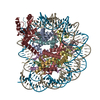
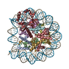
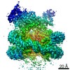
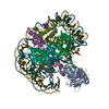
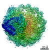
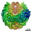
| Title | Structural model of 53BP1 bound to a ubiquitylated and methylated nucleosome, at 4.5 A resolution | ||||||||||||
 Components Components |
| ||||||||||||
 Keywords Keywords | STRUCTURAL PROTEIN/DNA / DNA / chromatin / 53BP1 / STRUCTURAL PROTEIN-DNA complex | ||||||||||||
| Function / homology |  Function and homology information Function and homology informationhistone H4K20me methyltransferase activity / positive regulation of isotype switching / ubiquitin-modified histone reader activity / histone H4K20me2 reader activity / cellular response to X-ray / double-strand break repair via classical nonhomologous end joining / protein localization to site of double-strand break / DNA repair complex / hypothalamus gonadotrophin-releasing hormone neuron development / female meiosis I ...histone H4K20me methyltransferase activity / positive regulation of isotype switching / ubiquitin-modified histone reader activity / histone H4K20me2 reader activity / cellular response to X-ray / double-strand break repair via classical nonhomologous end joining / protein localization to site of double-strand break / DNA repair complex / hypothalamus gonadotrophin-releasing hormone neuron development / female meiosis I / positive regulation of protein monoubiquitination / fat pad development / mitochondrion transport along microtubule / seminiferous tubule development / telomeric repeat DNA binding / female gonad development / SUMOylation of transcription factors / male meiosis I / histone reader activity / positive regulation of intrinsic apoptotic signaling pathway by p53 class mediator / negative regulation of double-strand break repair via homologous recombination / energy homeostasis / Replacement of protamines by nucleosomes in the male pronucleus / neuron projection morphogenesis / regulation of proteasomal protein catabolic process / Packaging Of Telomere Ends / Maturation of protein E / Recognition and association of DNA glycosylase with site containing an affected purine / Cleavage of the damaged purine / Maturation of protein E / ER Quality Control Compartment (ERQC) / Myoclonic epilepsy of Lafora / FLT3 signaling by CBL mutants / Deposition of new CENPA-containing nucleosomes at the centromere / IRAK2 mediated activation of TAK1 complex / Alpha-protein kinase 1 signaling pathway / Glycogen synthesis / IRAK1 recruits IKK complex / IRAK1 recruits IKK complex upon TLR7/8 or 9 stimulation / Prevention of phagosomal-lysosomal fusion / Endosomal Sorting Complex Required For Transport (ESCRT) / Membrane binding and targetting of GAG proteins / Negative regulation of FLT3 / Regulation of TBK1, IKKε (IKBKE)-mediated activation of IRF3, IRF7 / PTK6 Regulates RTKs and Their Effectors AKT1 and DOK1 / Regulation of TBK1, IKKε-mediated activation of IRF3, IRF7 upon TLR3 ligation / IRAK2 mediated activation of TAK1 complex upon TLR7/8 or 9 stimulation / Recognition and association of DNA glycosylase with site containing an affected pyrimidine / Cleavage of the damaged pyrimidine / Constitutive Signaling by NOTCH1 HD Domain Mutants / NOTCH2 Activation and Transmission of Signal to the Nucleus / TICAM1,TRAF6-dependent induction of TAK1 complex / DNA damage checkpoint signaling / TICAM1-dependent activation of IRF3/IRF7 / APC/C:Cdc20 mediated degradation of Cyclin B / RNA Polymerase I Promoter Opening / regulation of neuron apoptotic process / Downregulation of ERBB4 signaling / APC-Cdc20 mediated degradation of Nek2A / Regulation of FZD by ubiquitination / p75NTR recruits signalling complexes / Inhibition of DNA recombination at telomere / InlA-mediated entry of Listeria monocytogenes into host cells / TRAF6 mediated IRF7 activation in TLR7/8 or 9 signaling / Assembly of the ORC complex at the origin of replication / NF-kB is activated and signals survival / TRAF6-mediated induction of TAK1 complex within TLR4 complex / Regulation of pyruvate metabolism / Meiotic synapsis / Pexophagy / Downregulation of ERBB2:ERBB3 signaling / Regulation of innate immune responses to cytosolic DNA / NRIF signals cell death from the nucleus / Regulation of PTEN localization / Regulation of endogenous retroelements by the Human Silencing Hub (HUSH) complex / positive regulation of protein ubiquitination / replication fork / VLDLR internalisation and degradation / DNA methylation / Activated NOTCH1 Transmits Signal to the Nucleus / Condensation of Prophase Chromosomes / Synthesis of active ubiquitin: roles of E1 and E2 enzymes / Translesion synthesis by REV1 / Chromatin modifications during the maternal to zygotic transition (MZT) / SIRT1 negatively regulates rRNA expression / TICAM1, RIP1-mediated IKK complex recruitment / HCMV Late Events / Regulation of BACH1 activity / Translesion synthesis by POLK / InlB-mediated entry of Listeria monocytogenes into host cell / JNK (c-Jun kinases) phosphorylation and activation mediated by activated human TAK1 / MAP3K8 (TPL2)-dependent MAPK1/3 activation / Activation of IRF3, IRF7 mediated by TBK1, IKKε (IKBKE) / ERCC6 (CSB) and EHMT2 (G9a) positively regulate rRNA expression / Downregulation of TGF-beta receptor signaling / Translesion synthesis by POLI / Josephin domain DUBs / Gap-filling DNA repair synthesis and ligation in GG-NER / PRC2 methylates histones and DNA / IKK complex recruitment mediated by RIP1 Similarity search - Function | ||||||||||||
| Biological species |  Homo sapiens (human) Homo sapiens (human)synthetic construct (others) | ||||||||||||
| Method | ELECTRON MICROSCOPY / single particle reconstruction / cryo EM / Resolution: 4.54 Å | ||||||||||||
 Authors Authors | Wilson, M.D. / Benlekbir, S. / Sicheri, F. / Rubinstein, J.L. / Durocher, D. | ||||||||||||
| Funding support |  Canada, 3items Canada, 3items
| ||||||||||||
 Citation Citation | Journal: Nature / Year: 2016 Title: The structural basis of modified nucleosome recognition by 53BP1. Abstract: DNA double-strand breaks (DSBs) elicit a histone modification cascade that controls DNA repair. This pathway involves the sequential ubiquitination of histones H1 and H2A by the E3 ubiquitin ligases ...DNA double-strand breaks (DSBs) elicit a histone modification cascade that controls DNA repair. This pathway involves the sequential ubiquitination of histones H1 and H2A by the E3 ubiquitin ligases RNF8 and RNF168, respectively. RNF168 ubiquitinates H2A on lysine 13 and lysine 15 (refs 7, 8) (yielding H2AK13ub and H2AK15ub, respectively), an event that triggers the recruitment of 53BP1 (also known as TP53BP1) to chromatin flanking DSBs. 53BP1 binds specifically to H2AK15ub-containing nucleosomes through a peptide segment termed the ubiquitination-dependent recruitment motif (UDR), which requires the simultaneous engagement of histone H4 lysine 20 dimethylation (H4K20me2) by its tandem Tudor domain. How 53BP1 interacts with these two histone marks in the nucleosomal context, how it recognizes ubiquitin, and how it discriminates between H2AK13ub and H2AK15ub is unknown. Here we present the electron cryomicroscopy (cryo-EM) structure of a dimerized human 53BP1 fragment bound to a H4K20me2-containing and H2AK15ub-containing nucleosome core particle (NCP-ubme) at 4.5 Å resolution. The structure reveals that H4K20me2 and H2AK15ub recognition involves intimate contacts with multiple nucleosomal elements including the acidic patch. Ubiquitin recognition by 53BP1 is unusual and involves the sandwiching of the UDR segment between ubiquitin and the NCP surface. The selectivity for H2AK15ub is imparted by two arginine fingers in the H2A amino-terminal tail, which straddle the nucleosomal DNA and serve to position ubiquitin over the NCP-bound UDR segment. The structure of the complex between NCP-ubme and 53BP1 reveals the basis of 53BP1 recruitment to DSB sites and illuminates how combinations of histone marks and nucleosomal elements cooperate to produce highly specific chromatin responses, such as those elicited following chromosome breaks. | ||||||||||||
| History |
|
- Structure visualization
| Movie |
Movie viewer |
|---|---|
| Structure viewer | Molecule: MolmilJmol/JSmol |
- Downloads & links
-Download
| PDBx/mmCIF format | 5kgf.cif.gz | 365.4 KB | Display | PDBx/mmCIF format |
|---|---|---|---|---|
| PDB format | pdb5kgf.ent.gz | 274.6 KB | Display | PDB format |
| PDBx/mmJSON format | 5kgf.json.gz | Tree view | PDBx/mmJSON format | |
| Others |  Other downloads Other downloads |
-Validation report
| Arichive directory | https://data.pdbj.org/pub/pdb/validation_reports/kg/5kgfftp://data.pdbj.org/pub/pdb/validation_reports/kg/5kgf | HTTPS FTP |
|---|
-Related structure data
| Related structure data | 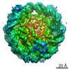 8246MC 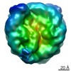 8247C M: map data used to model this data C: citing same article ( |
|---|---|
| Similar structure data |

-Links
 PDBj
PDBj


- Assembly
| Deposited unit | 
|
|---|---|
| 1 |
|
-Components
-Protein , 5 types, 10 molecules AEBFCGDHOM
| #1: Protein | Mass: 15421.101 Da / Num. of mol.: 2 Source method: isolated from a genetically manipulated source Source: (gene. exp.)  #2: Protein | Mass: 11439.511 Da / Num. of mol.: 2 / Mutation: K20C Source method: isolated from a genetically manipulated source Details: cysteine alkylation at position 20 / Source: (gene. exp.) #3: Protein | Mass: 14163.539 Da / Num. of mol.: 2 / Mutation: K13R, T16S, K36R Source method: isolated from a genetically manipulated source Details: Isopeptide amide crosslink between K15 of H2A and G76 of ubiquitin Source: (gene. exp.) Homo sapiens (human)Gene: HIST1H2AG, H2AFP, HIST1H2AI, H2AFC, HIST1H2AK, H2AFD, HIST1H2AL, H2AFI, HIST1H2AM, H2AFN Production host: #4: Protein | Mass: 13937.213 Da / Num. of mol.: 2 Source method: isolated from a genetically manipulated source Source: (gene. exp.) Homo sapiens (human)Gene: HIST1H2BC, H2BFL, HIST1H2BE, H2BFH, HIST1H2BF, H2BFG, HIST1H2BG, H2BFA, HIST1H2BI, H2BFK Production host: #8: Protein | Mass: 8576.831 Da / Num. of mol.: 2 Source method: isolated from a genetically manipulated source Source: (gene. exp.) Homo sapiens (human) / Gene: UBB / Production host: |
|---|
-DNA chain , 2 types, 2 molecules IJ
| #5: DNA chain | Mass: 44520.383 Da / Num. of mol.: 1 Source method: isolated from a genetically manipulated source Source: (gene. exp.) synthetic construct (others) / Production host: |
|---|---|
| #6: DNA chain | Mass: 44991.660 Da / Num. of mol.: 1 Source method: isolated from a genetically manipulated source Source: (gene. exp.) synthetic construct (others) / Production host: |
-Protein/peptide , 1 types, 2 molecules LK
| #7: Protein/peptide | Mass: 2376.757 Da / Num. of mol.: 2 Source method: isolated from a genetically manipulated source Details: full protein not modeled / Source: (gene. exp.) Homo sapiens (human) / Gene: TP53BP1 / Production host: |
|---|
-Details
| Has protein modification | Y |
|---|
-Experimental details
-Experiment
| Experiment | Method: ELECTRON MICROSCOPY |
|---|---|
| EM experiment | Aggregation state: PARTICLE / 3D reconstruction method: single particle reconstruction |
- Sample preparation
| Component |
| |||||||||||||||||||||||||||||||||||||||||||||||||||||||||||||||||||||||||||||
|---|---|---|---|---|---|---|---|---|---|---|---|---|---|---|---|---|---|---|---|---|---|---|---|---|---|---|---|---|---|---|---|---|---|---|---|---|---|---|---|---|---|---|---|---|---|---|---|---|---|---|---|---|---|---|---|---|---|---|---|---|---|---|---|---|---|---|---|---|---|---|---|---|---|---|---|---|---|---|
| Molecular weight |
| |||||||||||||||||||||||||||||||||||||||||||||||||||||||||||||||||||||||||||||
| Source (natural) |
| |||||||||||||||||||||||||||||||||||||||||||||||||||||||||||||||||||||||||||||
| Source (recombinant) |
| |||||||||||||||||||||||||||||||||||||||||||||||||||||||||||||||||||||||||||||
| Buffer solution | pH: 7.5 Details: High concentration NCP-ubme/GST-53BP1 complex at 200 mM salt was diluted just prior to grid freezing. | |||||||||||||||||||||||||||||||||||||||||||||||||||||||||||||||||||||||||||||
| Buffer component |
| |||||||||||||||||||||||||||||||||||||||||||||||||||||||||||||||||||||||||||||
| Specimen | Conc.: 0.6 mg/ml / Embedding applied: NO / Shadowing applied: NO / Staining applied: NO / Vitrification applied: YES | |||||||||||||||||||||||||||||||||||||||||||||||||||||||||||||||||||||||||||||
| Specimen support | Grid material: COPPER/RHODIUM / Grid mesh size: 400 divisions/in. / Grid type: electron micrsocopy sciences | |||||||||||||||||||||||||||||||||||||||||||||||||||||||||||||||||||||||||||||
| Vitrification | Instrument: FEI VITROBOT MARK III / Cryogen name: ETHANE-PROPANE / Humidity: 100 % / Chamber temperature: 277 K Details: Plunged into liquid ethane-propane (FEI VITROBOT MARK III) |
- Electron microscopy imaging
| Experimental equipment |  Model: Tecnai F20 / Image courtesy: FEI Company |
|---|---|
| Microscopy | Model: FEI TECNAI F20 |
| Electron gun | Electron source:  FIELD EMISSION GUN / Accelerating voltage: 200 kV / Illumination mode: FLOOD BEAM FIELD EMISSION GUN / Accelerating voltage: 200 kV / Illumination mode: FLOOD BEAM |
| Electron lens | Mode: BRIGHT FIELD / Nominal magnification: 25000 X / Calibrated magnification: 34483 X / Cs: 2 mm / C2 aperture diameter: 30 µm / Alignment procedure: COMA FREE |
| Specimen holder | Cryogen: NITROGEN Specimen holder model: GATAN 626 SINGLE TILT LIQUID NITROGEN CRYO TRANSFER HOLDER |
| Image recording | Average exposure time: 0.5 sec. / Electron dose: 36 e/Å2 / Detector mode: COUNTING / Film or detector model: GATAN K2 SUMMIT (4k x 4k) / Num. of grids imaged: 2 / Num. of real images: 319 |
| Image scans | Movie frames/image: 30 / Used frames/image: 1-30 |
- Processing
| Software | Name: PHENIX / Version: 1.10.1_2155: / Classification: refinement | ||||||||||||||||||||||||||||||||||||
|---|---|---|---|---|---|---|---|---|---|---|---|---|---|---|---|---|---|---|---|---|---|---|---|---|---|---|---|---|---|---|---|---|---|---|---|---|---|
| EM software |
| ||||||||||||||||||||||||||||||||||||
| CTF correction | Type: PHASE FLIPPING AND AMPLITUDE CORRECTION | ||||||||||||||||||||||||||||||||||||
| Particle selection | Num. of particles selected: 174185 Details: Automatically picked from roughly 3000 particles using a manually picked template | ||||||||||||||||||||||||||||||||||||
| Symmetry | Point symmetry: C2 (2 fold cyclic) | ||||||||||||||||||||||||||||||||||||
| 3D reconstruction | Resolution: 4.54 Å / Resolution method: FSC 0.143 CUT-OFF / Num. of particles: 45361 / Algorithm: FOURIER SPACE / Num. of class averages: 9 / Symmetry type: POINT | ||||||||||||||||||||||||||||||||||||
| Atomic model building | B value: 207.5 / Protocol: RIGID BODY FIT / Space: REAL Details: The atomic models of Widom-601 DNA (PDB ID 3LZ0), octameric histones (PDB ID 1KX5), ubiquitin (PDB ID 1UBI), and H4K20me2/53BP1 tandem Tudor domain (PDB ID 2IG0) were fitted without allowing ...Details: The atomic models of Widom-601 DNA (PDB ID 3LZ0), octameric histones (PDB ID 1KX5), ubiquitin (PDB ID 1UBI), and H4K20me2/53BP1 tandem Tudor domain (PDB ID 2IG0) were fitted without allowing flexibility into the 3D maps using UCSF Chimera. Segmentation was performed in UCSF Chimera. For the NCP-ubme structure the ubiquitin segmentation was further modified to remove obvious NCP density from the ubiquitin segment. The H2A/H2B sequence was mutated to the human H2AK13R/K36R and H2B manually in UCSF Chimera. A polyalanine model of the UDR was built within the UDR density in Coot, which compared well to predicted structures generated by Rosetta. The UDR model was mutated and fitted using UCSF Chimera, followed by iterative rounds of real-space refinement in PHENIX and model optimization in Coot. All figures were prepared in UCSF Chimera. | ||||||||||||||||||||||||||||||||||||
| Refinement | Highest resolution: 4.54 Å | ||||||||||||||||||||||||||||||||||||
| Refine LS restraints |
|
