 Movie
Movie Controller


+ Open data

- Basic information
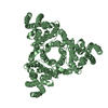
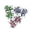
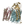
| Entry | Database: PDB / ID: 5kbj | ||||||
|---|---|---|---|---|---|---|---|
| Title | Structure of Rep-DNA complex | ||||||
 Components Components |
| ||||||
 Keywords Keywords | TRANSCRIPTION/DNA / Replication initiation / Rep protein / S. aureus / TRANSCRIPTION-DNA complex | ||||||
| Function / homology | Replication initiator A, N-terminal / Replication initiator protein A, C-terminal domain / Replication initiator protein A (RepA) N-terminus / Replication initiator protein A C-terminal domain / DNA / DNA (> 10) / Replication initiator A, N-terminal Function and homology information Function and homology information | ||||||
| Biological species |   Staphylococcus aureus (bacteria) Staphylococcus aureus (bacteria)synthetic construct (others) | ||||||
| Method |  X-RAY DIFFRACTION / SYNCHROTRON / MOLECULAR REPLACEMENT / Resolution: 3.09 Å X-RAY DIFFRACTION / SYNCHROTRON / MOLECULAR REPLACEMENT / Resolution: 3.09 Å | ||||||
 Authors Authors | Schumacher, M. | ||||||
 Citation Citation | Journal: Proc. Natl. Acad. Sci. U.S.A. / Year: 2014 Title: Mechanism of staphylococcal multiresistance plasmid replication origin assembly by the RepA protein. Authors: Schumacher, M.A. / Tonthat, N.K. / Kwong, S.M. / Chinnam, N.B. / Liu, M.A. / Skurray, R.A. / Firth, N. | ||||||
| History |
|
- Structure visualization
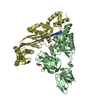
| Structure viewer | Molecule: MolmilJmol/JSmol |
|---|
- Downloads & links
-Download
| PDBx/mmCIF format | 5kbj.cif.gz | 258.6 KB | Display | PDBx/mmCIF format |
|---|---|---|---|---|
| PDB format | pdb5kbj.ent.gz | 205.8 KB | Display | PDB format |
| PDBx/mmJSON format | 5kbj.json.gz | Tree view | PDBx/mmJSON format | |
| Others |  Other downloads Other downloads |
-Validation report
| Summary document | 5kbj_validation.pdf.gz | 493.5 KB | Display | wwPDB validaton report |
|---|---|---|---|---|
| Full document | 5kbj_full_validation.pdf.gz | 531.9 KB | Display | |
| Data in XML | 5kbj_validation.xml.gz | 41.4 KB | Display | |
| Data in CIF | 5kbj_validation.cif.gz | 55.8 KB | Display | |
| Arichive directory | https://data.pdbj.org/pub/pdb/validation_reports/kb/5kbjftp://data.pdbj.org/pub/pdb/validation_reports/kb/5kbj | HTTPS FTP |
-Related structure data
| Related structure data |  4pqkC 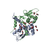 4pqlSC  4pt7C  4ptaC C: citing same article ( S: Starting model for refinement |
|---|---|
| Similar structure data |


-Links
 PDBj
PDBj


- Assembly
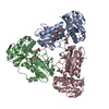
| Deposited unit | 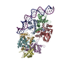
| ||||||||
|---|---|---|---|---|---|---|---|---|---|
| 1 | 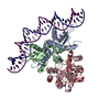
| ||||||||
| 2 | 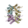
| ||||||||
| Unit cell |
|
-Components
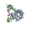
| #1: Protein | Mass: 15966.347 Da / Num. of mol.: 8 / Fragment: UNP residues 2-133 Source method: isolated from a genetically manipulated source Source: (gene. exp.) Staphylococcus aureus (bacteria) / Gene: SAP042A_013, SAP058A_012, SAP071A_014 / Production host: #2: DNA chain | | Mass: 9843.370 Da / Num. of mol.: 1 / Source method: obtained synthetically / Source: (synth.) synthetic construct (others) #3: DNA chain | | Mass: 9838.323 Da / Num. of mol.: 1 / Source method: obtained synthetically / Source: (synth.) synthetic construct (others) |
|---|
-Experimental details
-Experiment
| Experiment | Method: X-RAY DIFFRACTION / Number of used crystals: 1 |
|---|
- Sample preparation
| Crystal | Density Matthews: 2.87 Å3/Da / Density % sol: 57.18 % |
|---|---|
| Crystal grow | Temperature: 298 K / Method: vapor diffusion, hanging drop Details: 28% PEG 400, 0.1 M sodium/potassium phosphate (pH 6.9), 4% PEG 3350, and 10% polyethylene glycol |
-Data collection
| Diffraction | Mean temperature: 100 K |
|---|---|
| Diffraction source | Source: SYNCHROTRON / Site: ALS  / Beamline: 8.3.1 / Wavelength: 1 Å / Beamline: 8.3.1 / Wavelength: 1 Å |
| Detector | Type: ADSC QUANTUM 315r / Detector: CCD / Date: Feb 24, 2011 |
| Radiation | Protocol: SINGLE WAVELENGTH / Monochromatic (M) / Laue (L): M / Scattering type: x-ray |
| Radiation wavelength | Wavelength: 1 Å / Relative weight: 1 |
| Reflection | Resolution: 3.09→97.1 Å / Num. obs: 28484 / % possible obs: 94.5 % / Redundancy: 1.8 % / Net I/σ(I): 7 |
- Processing
| Software |
| |||||||||||||||||||||||||||||||||||||||||||||||||||||||||||||||||||||||||||||
|---|---|---|---|---|---|---|---|---|---|---|---|---|---|---|---|---|---|---|---|---|---|---|---|---|---|---|---|---|---|---|---|---|---|---|---|---|---|---|---|---|---|---|---|---|---|---|---|---|---|---|---|---|---|---|---|---|---|---|---|---|---|---|---|---|---|---|---|---|---|---|---|---|---|---|---|---|---|---|
| Refinement | Method to determine structure: MOLECULAR REPLACEMENT Starting model: 4PQL Resolution: 3.09→97.1 Å / SU ML: 0.46 / Cross valid method: FREE R-VALUE / σ(F): 1.98 / Phase error: 32.49
| |||||||||||||||||||||||||||||||||||||||||||||||||||||||||||||||||||||||||||||
| Solvent computation | Shrinkage radii: 0.27 Å / VDW probe radii: 0.6 Å / Bsol: 50.273 Å2 / ksol: 0.329 e/Å3 | |||||||||||||||||||||||||||||||||||||||||||||||||||||||||||||||||||||||||||||
| Displacement parameters | Biso max: 174.19 Å2 / Biso mean: 88.46 Å2 / Biso min: 30.68 Å2
| |||||||||||||||||||||||||||||||||||||||||||||||||||||||||||||||||||||||||||||
| Refinement step | Cycle: final / Resolution: 3.09→97.1 Å
| |||||||||||||||||||||||||||||||||||||||||||||||||||||||||||||||||||||||||||||
| Refine LS restraints |
| |||||||||||||||||||||||||||||||||||||||||||||||||||||||||||||||||||||||||||||
| LS refinement shell | Refine-ID: X-RAY DIFFRACTION / Total num. of bins used: 10
|
