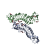
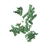
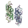
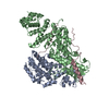
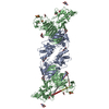
[English] 日本語
Yorodumi
- PDB-5k6k: Zika virus non-structural protein 1 (NS1) -

+
Open data


ID or keywords:

Loading...

-
Basic information

Entry
Database: PDB / ID: 5k6k
TitleZika virus non-structural protein 1 (NS1)
ComponentsZika virus protein
KeywordsVIRAL PROTEIN
Function / homology
Function and homology information


symbiont-mediated suppression of host JAK-STAT cascade via inhibition of host TYK2 activity / flavivirin / symbiont-mediated suppression of host JAK-STAT cascade via inhibition of STAT2 activity / symbiont-mediated suppression of host JAK-STAT cascade via inhibition of STAT1 activity / viral capsid / nucleoside-triphosphate phosphatase / double-stranded RNA binding / symbiont-mediated suppression of host cytoplasmic pattern recognition receptor signaling pathway via inhibition of TBK1 activity / symbiont-mediated suppression of host toll-like receptor signaling pathway / mRNA (guanine-N7)-methyltransferase ...symbiont-mediated suppression of host JAK-STAT cascade via inhibition of host TYK2 activity / flavivirin / symbiont-mediated suppression of host JAK-STAT cascade via inhibition of STAT2 activity / symbiont-mediated suppression of host JAK-STAT cascade via inhibition of STAT1 activity / viral capsid / nucleoside-triphosphate phosphatase / double-stranded RNA binding / symbiont-mediated suppression of host cytoplasmic pattern recognition receptor signaling pathway via inhibition of TBK1 activity / symbiont-mediated suppression of host toll-like receptor signaling pathway / mRNA (guanine-N7)-methyltransferase / methyltransferase cap1 / clathrin-dependent endocytosis of virus by host cell / mRNA (nucleoside-2'-O-)-methyltransferase activity / mRNA 5'-cap (guanine-N7-)-methyltransferase activity / RNA helicase activity / host cell endoplasmic reticulum membrane / host cell perinuclear region of cytoplasm / protein dimerization activity / RNA helicase / induction by virus of host autophagy / RNA-directed RNA polymerase / viral RNA genome replication / RNA-dependent RNA polymerase activity / serine-type endopeptidase activity / fusion of virus membrane with host endosome membrane / viral envelope / lipid binding / symbiont-mediated suppression of host type I interferon-mediated signaling pathway / host cell nucleus / structural molecule activity / virion attachment to host cell / GTP binding / virion membrane / ATP hydrolysis activity / proteolysis / extracellular region / ATP binding / membrane / metal ion binding
Similarity search - Function
: / : / Flavivirus envelope glycoprotein E, stem/anchor domain / RNA-directed RNA polymerase, thumb domain, Flavivirus / Flavivirus RNA-directed RNA polymerase, thumb domain / Flavivirus capsid protein C superfamily / Flavivirus non-structural protein NS2B / Flavivirus NS3 helicase, C-terminal helical domain / Genome polyprotein, Flavivirus / Flavivirus non-structural protein NS4A ...: / : / Flavivirus envelope glycoprotein E, stem/anchor domain / RNA-directed RNA polymerase, thumb domain, Flavivirus / Flavivirus RNA-directed RNA polymerase, thumb domain / Flavivirus capsid protein C superfamily / Flavivirus non-structural protein NS2B / Flavivirus NS3 helicase, C-terminal helical domain / Genome polyprotein, Flavivirus / Flavivirus non-structural protein NS4A / Flavivirus non-structural protein NS2B / Flavivirus capsid protein C / Flavivirus non-structural protein NS4B / mRNA cap 0/1 methyltransferase / Flavivirus capsid protein C / Flavivirus non-structural protein NS4B / Flavivirus non-structural protein NS4A / Flavivirus NS2B domain profile. / mRNA cap 0 and cap 1 methyltransferase (EC 2.1.1.56 and EC 2.1.1.57) domain profile. / Flavivirus non-structural protein NS2A / Flavivirus non-structural protein NS2A / Flavivirus NS3, petidase S7 / Peptidase S7, Flavivirus NS3 serine protease / Flavivirus NS3 protease (NS3pro) domain profile. / Envelope glycoprotein M, flavivirus / Flavivirus envelope glycoprotein M / RNA-directed RNA polymerase, flavivirus / Flavivirus RNA-directed RNA polymerase, fingers and palm domains / Flavivirus non-structural Protein NS1 / Flavivirus non-structural protein NS1 / Envelope glycoprotein M superfamily, flavivirus / Flavivirus polyprotein propeptide / Flavivirus polyprotein propeptide superfamily / Flavivirus polyprotein propeptide / Flaviviral glycoprotein E, central domain, subdomain 1 / Flaviviral glycoprotein E, central domain, subdomain 2 / Flavivirus envelope glycoprotein E, Stem/Anchor domain / Flavivirus glycoprotein E, immunoglobulin-like domain / Flavivirus envelope glycoprotein E, Stem/Anchor domain superfamily / Flavivirus glycoprotein, immunoglobulin-like domain / Flavivirus glycoprotein central and dimerisation domain / Flavivirus glycoprotein, central and dimerisation domains / Ribosomal RNA methyltransferase, FtsJ domain / FtsJ-like methyltransferase / Flavivirus/Alphavirus glycoprotein, immunoglobulin-like domain superfamily / Flavivirus glycoprotein, central and dimerisation domain superfamily / Flaviviral glycoprotein E, dimerisation domain / DEAD box, Flavivirus / Flavivirus DEAD domain / helicase superfamily c-terminal domain / Immunoglobulin E-set / Superfamilies 1 and 2 helicase C-terminal domain profile. / Superfamilies 1 and 2 helicase ATP-binding type-1 domain profile. / DEAD-like helicases superfamily / Helicase, C-terminal / Helicase superfamily 1/2, ATP-binding domain / RNA-directed RNA polymerase, catalytic domain / RdRp of positive ssRNA viruses catalytic domain profile. / S-adenosyl-L-methionine-dependent methyltransferase superfamily / Peptidase S1, PA clan / DNA/RNA polymerase superfamily / P-loop containing nucleoside triphosphate hydrolase
Similarity search - Domain/homology
Biological speciesZika virus
MethodX-RAY DIFFRACTION / SYNCHROTRON / MOLECULAR REPLACEMENT / molecular replacement / Resolution: 1.89 Å
AuthorsAkey, D.L. / Brown, W.C. / Smith, J.L.
CitationJournal: Nat.Struct.Mol.Biol. / Year: 2016
Title: Extended surface for membrane association in Zika virus NS1 structure.
Authors: Brown, W.C. / Akey, D.L. / Konwerski, J.R. / Tarrasch, J.T. / Skiniotis, G. / Kuhn, R.J. / Smith, J.L.
History
DepositionMay 24, 2016Deposition site: RCSB / Processing site: RCSB
Revision 1.0Jul 6, 2016Provider: repository / Type: Initial release
Revision 1.1Jul 13, 2016Group: Database references / Structure summary
Revision 1.2Aug 10, 2016Group: Database references
Revision 1.3Sep 21, 2016Group: Database references
Revision 1.4Nov 22, 2017Group: Derived calculations / Refinement description / Category: pdbx_struct_oper_list / software
Item: _pdbx_struct_oper_list.symmetry_operation / _software.classification
Revision 1.5Jul 29, 2020Group: Data collection / Derived calculations / Structure summary
Category: chem_comp / entity ...chem_comp / entity / pdbx_chem_comp_identifier / pdbx_entity_nonpoly / struct_conn / struct_site / struct_site_gen
Item: _chem_comp.name / _chem_comp.type ..._chem_comp.name / _chem_comp.type / _entity.pdbx_description / _pdbx_entity_nonpoly.name / _struct_conn.pdbx_role
Description: Carbohydrate remediation / Provider: repository / Type: Remediation
Revision 1.6Sep 27, 2023Group: Data collection / Database references ...Data collection / Database references / Refinement description / Structure summary
Category: chem_comp / chem_comp_atom ...chem_comp / chem_comp_atom / chem_comp_bond / database_2 / pdbx_initial_refinement_model
Item: _chem_comp.pdbx_synonyms / _database_2.pdbx_DOI / _database_2.pdbx_database_accession

-
Structure visualization

Structure viewerMolecule:
MolmilJmol/JSmol

Downloads & links

-
Assembly

Deposited unit
A: Zika virus protein
B: Zika virus protein
hetero molecules


Theoretical massNumber of molelcules
Total (without water)87,28014
Polymers85,6382
Non-polymers1,64112
Water18,8621047
1


  • Idetical with deposited unit
  • defined by author&software
TypeNameSymmetry operationNumber
identity operation1_555x,y,z1
Buried area7690 Å2
ΔGint-59 kcal/mol
Surface area31630 Å2
MethodPISA
2
A: Zika virus protein
B: Zika virus protein
hetero molecules

A: Zika virus protein
B: Zika virus protein
hetero molecules


Theoretical massNumber of molelcules
Total (without water)174,56028
Polymers171,2774
Non-polymers3,28324
Water724
TypeNameSymmetry operationNumber
identity operation1_555x,y,z1
crystal symmetry operation2_445-x-1,-y-1,z1
Buried area18120 Å2
ΔGint-133 kcal/mol
Surface area60510 Å2
MethodPISA
3
A: Zika virus protein
B: Zika virus protein
hetero molecules

A: Zika virus protein
B: Zika virus protein
hetero molecules


Theoretical massNumber of molelcules
Total (without water)174,56028
Polymers171,2774
Non-polymers3,28324
Water724
TypeNameSymmetry operationNumber
identity operation1_555x,y,z1
crystal symmetry operation3_555-x,y,-z1
Buried area17800 Å2
ΔGint-117 kcal/mol
Surface area60830 Å2
MethodPISA
Unit cell
Length a, b, c (Å)96.160, 130.440, 147.950
Angle α, β, γ (deg.)90.000, 90.000, 90.000
Int Tables number23
Space group name H-MI222
Components on special symmetry positions
IDModelComponents
11A-563-

HOH

21A-950-

HOH

-
Components

#1: Protein Zika virus protein /


Mass: 42819.164 Da / Num. of mol.: 2
Source method: isolated from a genetically manipulated source
Source: (gene. exp.) Zika virus / Strain: Mr 766 / Production host: unidentified baculovirus / References: UniProt: Q32ZE1
#2: Sugar
ChemComp-NAG / 2-acetamido-2-deoxy-beta-D-glucopyranose / N-acetyl-beta-D-glucosamine / 2-acetamido-2-deoxy-beta-D-glucose / 2-acetamido-2-deoxy-D-glucose / 2-acetamido-2-deoxy-glucose / N-ACETYL-D-GLUCOSAMINE / N-Acetylglucosamine


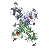
Type: D-saccharide, beta linking / Mass: 221.208 Da / Num. of mol.: 4
Source method: isolated from a genetically manipulated source
Formula: C8H15NO6
IdentifierTypeProgram
DGlcpNAcbCONDENSED IUPAC CARBOHYDRATE SYMBOLGMML 1.0
N-acetyl-b-D-glucopyranosamineCOMMON NAMEGMML 1.0
b-D-GlcpNAcIUPAC CARBOHYDRATE SYMBOLPDB-CARE 1.0
GlcNAcSNFG CARBOHYDRATE SYMBOLGMML 1.0
#3: Chemical
ChemComp-SO4 / SULFATE ION / Sulfate


Mass: 96.063 Da / Num. of mol.: 5 / Source method: obtained synthetically / Formula: SO4
#4: Chemical ChemComp-GOL / GLYCEROL / GLYCERIN / PROPANE-1,2,3-TRIOL / Glycerol


Mass: 92.094 Da / Num. of mol.: 3 / Source method: obtained synthetically / Formula: C3H8O3
#5: Water ChemComp-HOH / water / Water


Mass: 18.015 Da / Num. of mol.: 1047 / Source method: isolated from a natural source / Formula: H2O

-
Experimental details

-
Experiment

ExperimentMethod: X-RAY DIFFRACTION / Number of used crystals: 1

-
Sample preparation

CrystalDensity Matthews: 2.71 Å3/Da / Density % sol: 54.59 %
Crystal growTemperature: 293 K / Method: vapor diffusion, hanging drop / pH: 8.5 / Details: 25% PEG 3350 150 mM NH3SO4 100 mM tris 8.5

-
Data collection

DiffractionMean temperature: 100 K
Diffraction sourceSource: SYNCHROTRON / Site: APS / Beamline: 23-ID-D / Wavelength: 1.0332 Å
DetectorType: DECTRIS PILATUS3 6M / Detector: PIXEL / Date: Apr 21, 2016
RadiationMonochromator: Double Crystal / Protocol: SINGLE WAVELENGTH / Monochromatic (M) / Laue (L): M / Scattering type: x-ray
Radiation wavelengthWavelength: 1.0332 Å / Relative weight: 1
ReflectionResolution: 1.89→48.08 Å / Num. obs: 74493 / % possible obs: 100 % / Observed criterion σ(I): -3 / Redundancy: 13.5 % / Biso Wilson estimate: 32.05 Å2 / CC1/2: 0.999 / Rmerge(I) obs: 0.125 / Net I/σ(I): 13.22
Reflection shell
Resolution (Å)Highest resolution (Å)Redundancy (%)Rmerge(I) obsMean I/σ(I) obsDiffraction-ID% possible all
1.89-1.9213.31.6131.331100
1.92-1.961.4611.61100
1.96-21.1092.161100
2-2.040.9342.621100
2.04-2.080.7983.11100
2.08-2.130.6693.671100
2.13-2.180.5664.531100
2.18-2.240.5084.981100
2.24-2.310.4275.971100
2.31-2.380.357.211100
2.38-2.470.2918.541100
2.47-2.570.23910.11100
2.57-2.680.19712.31100
2.68-2.830.15614.931100
2.83-30.12419.041100
3-3.230.123.191100
3.23-3.560.07928.251100
3.56-4.080.06833.921100
4.08-5.130.06236.981100
5.130.0637.29199.8

-
Phasing

PhasingMethod: molecular replacement
Phasing MR
Highest resolutionLowest resolution
Rotation7.07 Å48.61 Å
Translation7.07 Å48.61 Å

-
Processing

Software
NameVersionClassification
XSCALEdata scaling
XDSdata scaling
PHASER2.6.0phasing
PHENIXrefinement
PDB_EXTRACT3.2data extraction
RefinementMethod to determine structure: MOLECULAR REPLACEMENT
Starting model: 4O6D
Resolution: 1.89→48.08 Å / SU ML: 0.24 / Cross valid method: FREE R-VALUE / σ(F): 1.35 / Phase error: 18.92
RfactorNum. reflection% reflection
Rfree0.1889 2089 2.81 %
Rwork0.1486 --
obs0.1498 74473 99.97 %
Solvent computationShrinkage radii: 0.9 Å / VDW probe radii: 1.11 Å
Displacement parametersBiso max: 139.22 Å2 / Biso mean: 38.6056 Å2 / Biso min: 15.11 Å2
Refinement stepCycle: final / Resolution: 1.89→48.08 Å
ProteinNucleic acidLigandSolventTotal
Num. atoms5624 0 99 1047 6770
Biso mean--80.31 51.33 -
Num. residues----704
Refine LS restraints
Refine-IDTypeDev idealNumber
X-RAY DIFFRACTIONf_bond_d0.0075933
X-RAY DIFFRACTIONf_angle_d0.8388069
X-RAY DIFFRACTIONf_chiral_restr0.055850
X-RAY DIFFRACTIONf_plane_restr0.0051035
X-RAY DIFFRACTIONf_dihedral_angle_d18.0033539
LS refinement shell

Refine-ID: X-RAY DIFFRACTION / Total num. of bins used: 15 / % reflection obs: 100 %

Resolution (Å)Rfactor RfreeNum. reflection RfreeRfactor RworkNum. reflection RworkNum. reflection all
1.8901-1.9340.35981360.30547694905
1.934-1.98240.31561400.255747634903
1.9824-2.0360.25491420.212947804922
2.036-2.09590.23591370.187247724909
2.0959-2.16360.23411340.174347814915
2.1636-2.24090.18391390.170448284967
2.2409-2.33060.18831390.169747794918
2.3306-2.43670.22761370.157947994936
2.4367-2.56510.20551380.153748024940
2.5651-2.72580.1891370.153248284965
2.7258-2.93630.1891410.146647994940
2.9363-3.23170.18261400.138148675007
3.2317-3.69920.17561420.129148524994
3.6992-4.660.15921410.112748935034
4.66-48.09550.16411460.145650725218
Refinement TLS params.

Method: refined / Refine-ID: X-RAY DIFFRACTION

IDL112)L122)L132)L222)L232)L332)S11 (Å °)S12 (Å °)S13 (Å °)S21 (Å °)S22 (Å °)S23 (Å °)S31 (Å °)S32 (Å °)S33 (Å °)T112)T122)T132)T222)T232)T332)Origin x (Å)Origin y (Å)Origin z (Å)
12.81881.39070.39731.1370.00460.80990.0568-0.0841-0.26350.0821-0.0306-0.23430.02040.133-0.01990.21550.0236-0.03130.23110.0080.2535-3.2237-38.7751-14.1706
20.4647-0.3311-0.31122.10461.30832.0336-0.0044-0.06920.06850.00090.00250.0532-0.1672-0.0659-0.00060.1858-0.0099-0.01620.21590.02280.1965-27.1421-23.9272-11.8706
32.32460.27731.37542.56281.01644.29820.2611-0.2718-0.28380.1708-0.0963-0.14540.3299-0.3158-0.140.2462-0.0321-0.03280.18550.07080.2511-26.1711-55.0785-17.5835
42.96330.927-0.39272.5514-0.55911.72380.0845-0.2606-0.15450.1607-0.06780.25280.1387-0.106-0.01620.19-0.01720.00240.21180.02060.2252-56.8414-51.5902-31.0621
50.4988-0.1204-0.47831.05940.99213.629-0.0150.0514-0.0827-0.125-0.0131-0.04150.15190.13420.03380.22170.0008-0.0060.19040.02270.2229-26.294-51.0775-44.0288
Refinement TLS group
IDRefine-IDRefine TLS-IDSelection detailsAuth asym-IDAuth seq-ID
1X-RAY DIFFRACTION1chain 'A' and (resid -5 through 180 )A-5 - 180
2X-RAY DIFFRACTION2chain 'A' and (resid 181 through 352 )A181 - 352
3X-RAY DIFFRACTION3chain 'B' and (resid 0 through 31 )B0 - 31
4X-RAY DIFFRACTION4chain 'B' and (resid 32 through 180 )B32 - 180
5X-RAY DIFFRACTION5chain 'B' and (resid 181 through 352 )B181 - 352

+
About Yorodumi

-
News

-
Feb 9, 2022. New format data for meta-information of EMDB entries

New format data for meta-information of EMDB entries

  • Version 3 of the EMDB header file is now the official format.
  • The previous official version 1.9 will be removed from the archive.

Related info.:EMDB header

External links:wwPDB to switch to version 3 of the EMDB data model

-
Aug 12, 2020. Covid-19 info

Covid-19 info

URL: https://pdbj.org/emnavi/covid19.php

New page: Covid-19 featured information page in EM Navigator.

Related info.:Covid-19 info / Mar 5, 2020. Novel coronavirus structure data

+
Mar 5, 2020. Novel coronavirus structure data

Novel coronavirus structure data

Related info.:Yorodumi Speices / Aug 12, 2020. Covid-19 info

External links:COVID-19 featured content - PDBj / Molecule of the Month (242):Coronavirus Proteases

+
Jan 31, 2019. EMDB accession codes are about to change! (news from PDBe EMDB page)

EMDB accession codes are about to change! (news from PDBe EMDB page)

  • The allocation of 4 digits for EMDB accession codes will soon come to an end. Whilst these codes will remain in use, new EMDB accession codes will include an additional digit and will expand incrementally as the available range of codes is exhausted. The current 4-digit format prefixed with “EMD-” (i.e. EMD-XXXX) will advance to a 5-digit format (i.e. EMD-XXXXX), and so on. It is currently estimated that the 4-digit codes will be depleted around Spring 2019, at which point the 5-digit format will come into force.
  • The EM Navigator/Yorodumi systems omit the EMD- prefix.

Related info.:Q: What is EMD? / ID/Accession-code notation in Yorodumi/EM Navigator

External links:EMDB Accession Codes are Changing Soon! / Contact to PDBj

+
Jul 12, 2017. Major update of PDB

Major update of PDB

  • wwPDB released updated PDB data conforming to the new PDBx/mmCIF dictionary.
  • This is a major update changing the version number from 4 to 5, and with Remediation, in which all the entries are updated.
  • In this update, many items about electron microscopy experimental information are reorganized (e.g. em_software).
  • Now, EM Navigator and Yorodumi are based on the updated data.

External links:wwPDB Remediation / Enriched Model Files Conforming to OneDep Data Standards Now Available in the PDB FTP Archive

-
Yorodumi

Thousand views of thousand structures

  • Yorodumi is a browser for structure data from EMDB, PDB, SASBDB, etc.
  • This page is also the successor to EM Navigator detail page, and also detail information page/front-end page for Omokage search.
  • The word "yorodu" (or yorozu) is an old Japanese word meaning "ten thousand". "mi" (miru) is to see.

Related info.:EMDB / PDB / SASBDB / Comparison of 3 databanks / Yorodumi Search / Aug 31, 2016. New EM Navigator & Yorodumi / Yorodumi Papers / Jmol/JSmol / Function and homology information / Changes in new EM Navigator and Yorodumi

Read more