 Movie
Movie Controller

[English] 日本語
 Yorodumi
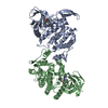
- PDB-5k5q: Structure of AspA-DNA complex: novel centromere bindng protein-ce... -
+ Open data

- Basic information
| Entry | Database: PDB / ID: 5k5q | |||||||||
|---|---|---|---|---|---|---|---|---|---|---|
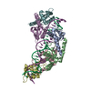
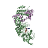
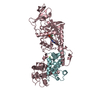
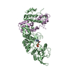
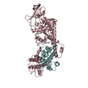
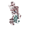
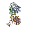
| Title | Structure of AspA-DNA complex: novel centromere bindng protein-centromere complex | |||||||||
 Components Components |
| |||||||||
 Keywords Keywords | TRANSCRIPTION/DNA / AspA / centromere / segregation / archaea / pNOB8 / TRANSCRIPTION-DNA complex | |||||||||
| Function / homology |  Function and homology information Function and homology information | |||||||||
| Biological species |   Sulfolobus sp. NOB8H2 (acidophilic) Sulfolobus sp. NOB8H2 (acidophilic)synthetic construct (others) | |||||||||
| Method |  X-RAY DIFFRACTION / SYNCHROTRON / MOLECULAR REPLACEMENT / molecular replacement / Resolution: 2.649 Å X-RAY DIFFRACTION / SYNCHROTRON / MOLECULAR REPLACEMENT / molecular replacement / Resolution: 2.649 Å | |||||||||
 Authors Authors | Schumacher, M. | |||||||||
 Citation Citation | Journal: Science / Year: 2015 Title: Structures of archaeal DNA segregation machinery reveal bacterial and eukaryotic linkages. Authors: Schumacher, M.A. / Tonthat, N.K. / Lee, J. / Rodriguez-Castaneda, F.A. / Chinnam, N.B. / Kalliomaa-Sanford, A.K. / Ng, I.W. / Barge, M.T. / Shaw, P.L. / Barilla, D. | |||||||||
| History |
|
- Structure visualization
| Structure viewer | Molecule: MolmilJmol/JSmol |
|---|
- Downloads & links
-Download
| PDBx/mmCIF format | 5k5q.cif.gz | 157.1 KB | Display | PDBx/mmCIF format |
|---|---|---|---|---|
| PDB format | pdb5k5q.ent.gz | 120.2 KB | Display | PDB format |
| PDBx/mmJSON format | 5k5q.json.gz | Tree view | PDBx/mmJSON format | |
| Others |  Other downloads Other downloads |
-Validation report
| Arichive directory | https://data.pdbj.org/pub/pdb/validation_reports/k5/5k5qftp://data.pdbj.org/pub/pdb/validation_reports/k5/5k5q | HTTPS FTP |
|---|
-Related structure data
| Related structure data | 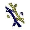 4rs7C  4rs8SC 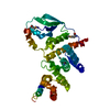 5k5aC 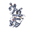 5k5dC  5k5oC 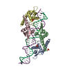 5k5rC 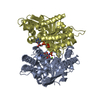 5k5zC S: Starting model for refinement C: citing same article ( |
|---|---|
| Similar structure data |


-Links
 PDBj
PDBj


- Assembly
| Deposited unit | 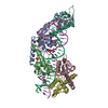
| ||||||||
|---|---|---|---|---|---|---|---|---|---|
| 1 |
| ||||||||
| Unit cell |
|
-Components
| #1: Protein | Mass: 10596.363 Da / Num. of mol.: 6 Source method: isolated from a genetically manipulated source Source: (gene. exp.) Sulfolobus sp. NOB8H2 (acidophilic) / Production host:  #2: DNA chain | | Mass: 9829.362 Da / Num. of mol.: 1 / Source method: obtained synthetically / Source: (synth.) synthetic construct (others) #3: DNA chain | | Mass: 9846.403 Da / Num. of mol.: 1 / Source method: obtained synthetically / Source: (synth.) synthetic construct (others) #4: Chemical |   Mass: 94.971 Da / Num. of mol.: 2 / Source method: obtained synthetically / Formula: PO4 Mass: 94.971 Da / Num. of mol.: 2 / Source method: obtained synthetically / Formula: PO4#5: Water | ChemComp-HOH / |  Mass: 18.015 Da / Num. of mol.: 5 / Source method: isolated from a natural source / Formula: H2O Mass: 18.015 Da / Num. of mol.: 5 / Source method: isolated from a natural source / Formula: H2O |
|---|
-Experimental details
-Experiment
| Experiment | Method: X-RAY DIFFRACTION / Number of used crystals: 1 |
|---|
- Sample preparation
| Crystal | Density Matthews: 2.54 Å3/Da / Density % sol: 51.55 % |
|---|---|
| Crystal grow | Temperature: 298 K / Method: vapor diffusion, hanging drop / pH: 6.2 Details: 10% PEG 3000, 0.1 M sodium phosphate/citrate pH 6.2 |
-Data collection
| Diffraction | Mean temperature: 100 K |
|---|---|
| Diffraction source | Source: SYNCHROTRON / Site: ALS  / Beamline: 8.3.1 / Wavelength: 1 Å / Beamline: 8.3.1 / Wavelength: 1 Å |
| Detector | Type: ADSC QUANTUM 315r / Detector: CCD / Date: Dec 20, 2011 |
| Radiation | Protocol: SINGLE WAVELENGTH / Monochromatic (M) / Laue (L): M / Scattering type: x-ray |
| Radiation wavelength | Wavelength: 1 Å / Relative weight: 1 |
| Reflection | Resolution: 2.649→48.018 Å / Num. obs: 22831 / % possible obs: 92.64 % / Redundancy: 4 % / Rsym value: 0.058 / Net I/σ(I): 8.5 |
-Phasing
| Phasing | Method: molecular replacement |
|---|
- Processing
| Software |
| |||||||||||||||||||||||||||||||||||||||||||||||||||||||||||||||||||||||||||||
|---|---|---|---|---|---|---|---|---|---|---|---|---|---|---|---|---|---|---|---|---|---|---|---|---|---|---|---|---|---|---|---|---|---|---|---|---|---|---|---|---|---|---|---|---|---|---|---|---|---|---|---|---|---|---|---|---|---|---|---|---|---|---|---|---|---|---|---|---|---|---|---|---|---|---|---|---|---|---|
| Refinement | Method to determine structure: MOLECULAR REPLACEMENT Starting model: 4RS8 Resolution: 2.649→48.018 Å / SU ML: 0.42 / Cross valid method: FREE R-VALUE / σ(F): 0 / Phase error: 39.32
| |||||||||||||||||||||||||||||||||||||||||||||||||||||||||||||||||||||||||||||
| Solvent computation | Shrinkage radii: 0.95 Å / VDW probe radii: 1.2 Å / Bsol: 60.354 Å2 / ksol: 0.314 e/Å3 | |||||||||||||||||||||||||||||||||||||||||||||||||||||||||||||||||||||||||||||
| Displacement parameters | Biso max: 174.12 Å2 / Biso mean: 100.29 Å2 / Biso min: 34.79 Å2
| |||||||||||||||||||||||||||||||||||||||||||||||||||||||||||||||||||||||||||||
| Refinement step | Cycle: final / Resolution: 2.649→48.018 Å
| |||||||||||||||||||||||||||||||||||||||||||||||||||||||||||||||||||||||||||||
| Refine LS restraints |
| |||||||||||||||||||||||||||||||||||||||||||||||||||||||||||||||||||||||||||||
| LS refinement shell | Refine-ID: X-RAY DIFFRACTION / Total num. of bins used: 10
|
