 Movie
Movie Controller


+ Open data

- Basic information
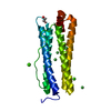
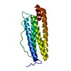
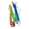
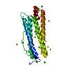
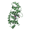
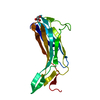
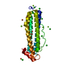
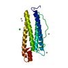
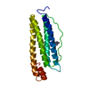
| Entry | Database: PDB / ID: 4rs7 | ||||||
|---|---|---|---|---|---|---|---|
| Title | Structure of pNOB8 ParB-C | ||||||
 Components Components | ParB-C | ||||||
 Keywords Keywords | DNA BINDING PROTEIN / histone-like fold / DNA segregation | ||||||
| Function / homology | : / Ssol_1539-like, second domain / ParB N-terminal domain / ParB/Sulfiredoxin / ParB-like nuclease domain / ParB/Sulfiredoxin superfamily / identical protein binding / ParB/Sulfiredoxin domain-containing protein Function and homology information Function and homology information | ||||||
| Biological species |   Sulfolobus sp. NOB8H2 (acidophilic) Sulfolobus sp. NOB8H2 (acidophilic) | ||||||
| Method |  X-RAY DIFFRACTION / SYNCHROTRON / MAD / Resolution: 1.9 Å X-RAY DIFFRACTION / SYNCHROTRON / MAD / Resolution: 1.9 Å | ||||||
 Authors Authors | Schumacher, M.A. / Lee, J. / Chinnam, N.B. / Barilla, D. | ||||||
 Citation Citation | Journal: Science / Year: 2015 Title: Structures of archaeal DNA segregation machinery reveal bacterial and eukaryotic linkages. Authors: Schumacher, M.A. / Tonthat, N.K. / Lee, J. / Rodriguez-Castaneda, F.A. / Chinnam, N.B. / Kalliomaa-Sanford, A.K. / Ng, I.W. / Barge, M.T. / Shaw, P.L. / Barilla, D. | ||||||
| History |
|
- Structure visualization
| Structure viewer | Molecule: MolmilJmol/JSmol |
|---|
- Downloads & links
-Download
| PDBx/mmCIF format | 4rs7.cif.gz | 92 KB | Display | PDBx/mmCIF format |
|---|---|---|---|---|
| PDB format | pdb4rs7.ent.gz | 71.5 KB | Display | PDB format |
| PDBx/mmJSON format | 4rs7.json.gz | Tree view | PDBx/mmJSON format | |
| Others |  Other downloads Other downloads |
-Validation report
| Arichive directory | https://data.pdbj.org/pub/pdb/validation_reports/rs/4rs7ftp://data.pdbj.org/pub/pdb/validation_reports/rs/4rs7 | HTTPS FTP |
|---|
-Related structure data
| Related structure data |  4rs8C 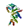 5k5aC 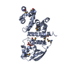 5k5dC  5k5oC 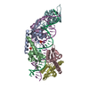 5k5qC 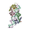 5k5rC 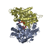 5k5zC C: citing same article ( |
|---|---|
| Similar structure data |

-Links
 PDBj
PDBj- Assembly
| Deposited unit | 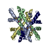
| ||||||||
|---|---|---|---|---|---|---|---|---|---|
| 1 | 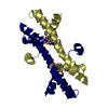
| ||||||||
| 2 | 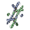
| ||||||||
| Unit cell |
|
-Components
| #1: Protein | Mass: 11725.104 Da / Num. of mol.: 4 / Fragment: UNP residues 375-470 Source method: isolated from a genetically manipulated source Source: (gene. exp.) Sulfolobus sp. NOB8H2 (acidophilic) / Production host:  #2: Water | ChemComp-HOH / |  Mass: 18.015 Da / Num. of mol.: 366 / Source method: isolated from a natural source / Formula: H2O Mass: 18.015 Da / Num. of mol.: 366 / Source method: isolated from a natural source / Formula: H2OHas protein modification | Y | |
|---|
-Experimental details
-Experiment
| Experiment | Method: X-RAY DIFFRACTION / Number of used crystals: 1 |
|---|
- Sample preparation
| Crystal | Density Matthews: 2.16 Å3/Da / Density % sol: 43.14 % |
|---|---|
| Crystal grow | Temperature: 298 K / Method: vapor diffusion, hanging drop / pH: 7.5 Details: PEG400, PEG8000, HEPES, pH 7.5, VAPOR DIFFUSION, HANGING DROP, temperature 298K |
-Data collection
| Diffraction | Mean temperature: 100 K | ||||||||||||
|---|---|---|---|---|---|---|---|---|---|---|---|---|---|
| Diffraction source | Source: SYNCHROTRON / Site: ALS  / Beamline: 8.3.1 / Wavelength: 0.979, 0.980, 0.9252 / Beamline: 8.3.1 / Wavelength: 0.979, 0.980, 0.9252 | ||||||||||||
| Detector | Type: ADSC QUANTUM 315r / Detector: CCD / Date: Jan 22, 2013 | ||||||||||||
| Radiation | Monochromator: double flat crystal Si(111) / Protocol: MAD / Monochromatic (M) / Laue (L): M / Scattering type: x-ray | ||||||||||||
| Radiation wavelength |
| ||||||||||||
| Reflection | Resolution: 1.9→37.34 Å / Num. all: 59766 / Num. obs: 59754 / % possible obs: 99 % / Observed criterion σ(F): 0 / Observed criterion σ(I): 0 | ||||||||||||
| Reflection shell | Highest resolution: 1.9 Å |
- Processing
| Software |
| |||||||||||||||||||||||||||||||||||||||||||||||||||||||||||||||||||||||||||||
|---|---|---|---|---|---|---|---|---|---|---|---|---|---|---|---|---|---|---|---|---|---|---|---|---|---|---|---|---|---|---|---|---|---|---|---|---|---|---|---|---|---|---|---|---|---|---|---|---|---|---|---|---|---|---|---|---|---|---|---|---|---|---|---|---|---|---|---|---|---|---|---|---|---|---|---|---|---|---|
| Refinement | Method to determine structure: MAD / Resolution: 1.9→37.34 Å / SU ML: 0.22 / σ(F): 0 / Phase error: 25.59 / Stereochemistry target values: ML
| |||||||||||||||||||||||||||||||||||||||||||||||||||||||||||||||||||||||||||||
| Solvent computation | Shrinkage radii: 0.9 Å / VDW probe radii: 1.11 Å / Solvent model: FLAT BULK SOLVENT MODEL / Bsol: 47.72 Å2 / ksol: 0.417 e/Å3 | |||||||||||||||||||||||||||||||||||||||||||||||||||||||||||||||||||||||||||||
| Displacement parameters |
| |||||||||||||||||||||||||||||||||||||||||||||||||||||||||||||||||||||||||||||
| Refinement step | Cycle: LAST / Resolution: 1.9→37.34 Å
| |||||||||||||||||||||||||||||||||||||||||||||||||||||||||||||||||||||||||||||
| Refine LS restraints |
| |||||||||||||||||||||||||||||||||||||||||||||||||||||||||||||||||||||||||||||
| LS refinement shell |
|
