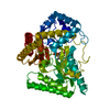
+Open data
-Basic information
Entry | Database: PDB / ID: 5jsi | ||||||
---|---|---|---|---|---|---|---|
Title | Structure of membrane protein | ||||||
Components | Bacteriorhodopsin | ||||||
Keywords | UNKNOWN FUNCTION / membrane protein / iodide / TRANSFERASE | ||||||
Function / homology | Bacteriorhodopsin-like protein / Archaeal/bacterial/fungal rhodopsins / Bacteriorhodopsin-like protein / membrane / IODIDE ION / EICOSANE / (2R)-2,3-dihydroxypropyl (9Z)-octadec-9-enoate / Bacteriorhodopsin Function and homology information | ||||||
Biological species | Candidatus Actinomarina minuta (bacteria) | ||||||
Method | X-RAY DIFFRACTION / SYNCHROTRON / SAD / Resolution: 2 Å | ||||||
Authors | Melnikov, I. / Polovinkin, V. / Kovalev, K. / Shevchenko, V. / Gushchin, I. / Popov, A. / Gordeliy, V. | ||||||
Citation | Journal: Sci Adv / Year: 2017 Title: Fast iodide-SAD phasing for high-throughput membrane protein structure determination. Authors: Melnikov, I. / Polovinkin, V. / Kovalev, K. / Gushchin, I. / Shevtsov, M. / Shevchenko, V. / Mishin, A. / Alekseev, A. / Rodriguez-Valera, F. / Borshchevskiy, V. / Cherezov, V. / Leonard, G. ...Authors: Melnikov, I. / Polovinkin, V. / Kovalev, K. / Gushchin, I. / Shevtsov, M. / Shevchenko, V. / Mishin, A. / Alekseev, A. / Rodriguez-Valera, F. / Borshchevskiy, V. / Cherezov, V. / Leonard, G.A. / Gordeliy, V. / Popov, A. | ||||||
History |
|
-Structure visualization
Structure viewer | Molecule: MolmilJmol/JSmol |
---|
-Downloads & links
-Download
PDBx/mmCIF format | 5jsi.cif.gz | 101.2 KB | Display | PDBx/mmCIF format |
---|---|---|---|---|
PDB format | pdb5jsi.ent.gz | 80.9 KB | Display | PDB format |
PDBx/mmJSON format | 5jsi.json.gz | Tree view | PDBx/mmJSON format | |
Others | Other downloads |
-Validation report
Summary document | 5jsi_validation.pdf.gz | 3.7 MB | Display | wwPDB validaton report |
---|---|---|---|---|
Full document | 5jsi_full_validation.pdf.gz | 3.7 MB | Display | |
Data in XML | 5jsi_validation.xml.gz | 21.2 KB | Display | |
Data in CIF | 5jsi_validation.cif.gz | 28.3 KB | Display | |
Arichive directory | https://data.pdbj.org/pub/pdb/validation_reports/js/5jsi ftp://data.pdbj.org/pub/pdb/validation_reports/js/5jsi | HTTPS FTP |
-Related structure data
-Links
-Assembly
Deposited unit |
| ||||||||
---|---|---|---|---|---|---|---|---|---|
1 |
| ||||||||
Unit cell |
|
-Components
#1: Protein | Mass: 24544.504 Da / Num. of mol.: 2 Source method: isolated from a genetically manipulated source Source: (gene. exp.) Candidatus Actinomarina minuta (bacteria) Production host: Escherichia coli (E. coli) / References: UniProt: S5DM51 #2: Chemical | ChemComp-LFA / #3: Chemical | ChemComp-IOD / #4: Chemical | ChemComp-OLC / ( | #5: Water | ChemComp-HOH / | |
---|
-Experimental details
-Experiment
Experiment | Method: X-RAY DIFFRACTION / Number of used crystals: 1 |
---|
-Sample preparation
Crystal | Density Matthews: 2.46 Å3/Da / Density % sol: 50.02 % |
---|---|
Crystal grow | Temperature: 293 K / Method: lipidic cubic phase / Details: in meso lipidic cubic phase crystallization |
-Data collection
Diffraction | Mean temperature: 100 K |
---|---|
Diffraction source | Source: SYNCHROTRON / Site: ESRF / Beamline: ID23-1 / Wavelength: 1.85 Å |
Detector | Type: DECTRIS PILATUS 6M-F / Detector: PIXEL / Date: Nov 10, 2015 |
Radiation | Protocol: SINGLE WAVELENGTH / Monochromatic (M) / Laue (L): M / Scattering type: x-ray |
Radiation wavelength | Wavelength: 1.85 Å / Relative weight: 1 |
Reflection | Resolution: 2→60 Å / Num. obs: 49905 / % possible obs: 81.9 % / Redundancy: 1.88 % / CC1/2: 0.997 / Rmerge(I) obs: 0.051 / Net I/σ(I): 8.64 |
Reflection shell | Resolution: 2→2.1 Å / Redundancy: 1.83 % / Rmerge(I) obs: 0.501 / Mean I/σ(I) obs: 1.43 / % possible all: 68.6 |
-Processing
Software |
| ||||||||||||||||||||||||||||||||||||||||||||||||||||||||||||||||||||||||||||||||||||||||||||||||||||||||||||||||||||||||||||||||||||||||||||||||||||||||||||||||||||||||||||||||||||||
---|---|---|---|---|---|---|---|---|---|---|---|---|---|---|---|---|---|---|---|---|---|---|---|---|---|---|---|---|---|---|---|---|---|---|---|---|---|---|---|---|---|---|---|---|---|---|---|---|---|---|---|---|---|---|---|---|---|---|---|---|---|---|---|---|---|---|---|---|---|---|---|---|---|---|---|---|---|---|---|---|---|---|---|---|---|---|---|---|---|---|---|---|---|---|---|---|---|---|---|---|---|---|---|---|---|---|---|---|---|---|---|---|---|---|---|---|---|---|---|---|---|---|---|---|---|---|---|---|---|---|---|---|---|---|---|---|---|---|---|---|---|---|---|---|---|---|---|---|---|---|---|---|---|---|---|---|---|---|---|---|---|---|---|---|---|---|---|---|---|---|---|---|---|---|---|---|---|---|---|---|---|---|---|
Refinement | Method to determine structure: SAD / Resolution: 2→50.8 Å / Cor.coef. Fo:Fc: 0.956 / Cor.coef. Fo:Fc free: 0.948 / SU B: 4.49 / SU ML: 0.124 / Cross valid method: THROUGHOUT / ESU R: 0.224 / ESU R Free: 0.179 / Stereochemistry target values: MAXIMUM LIKELIHOOD / Details: HYDROGENS HAVE BEEN ADDED IN THE RIDING POSITIONS
| ||||||||||||||||||||||||||||||||||||||||||||||||||||||||||||||||||||||||||||||||||||||||||||||||||||||||||||||||||||||||||||||||||||||||||||||||||||||||||||||||||||||||||||||||||||||
Solvent computation | Ion probe radii: 0.8 Å / Shrinkage radii: 0.8 Å / VDW probe radii: 1.2 Å / Solvent model: MASK | ||||||||||||||||||||||||||||||||||||||||||||||||||||||||||||||||||||||||||||||||||||||||||||||||||||||||||||||||||||||||||||||||||||||||||||||||||||||||||||||||||||||||||||||||||||||
Displacement parameters | Biso mean: 36.3 Å2
| ||||||||||||||||||||||||||||||||||||||||||||||||||||||||||||||||||||||||||||||||||||||||||||||||||||||||||||||||||||||||||||||||||||||||||||||||||||||||||||||||||||||||||||||||||||||
Refinement step | Cycle: 1 / Resolution: 2→50.8 Å
| ||||||||||||||||||||||||||||||||||||||||||||||||||||||||||||||||||||||||||||||||||||||||||||||||||||||||||||||||||||||||||||||||||||||||||||||||||||||||||||||||||||||||||||||||||||||
Refine LS restraints |
|