 Movie
Movie Controller

[English] 日本語
 Yorodumi
- PDB-5ftf: Crystal structure of Pif1 helicase from Bacteroides double mutant... -
+ Open data

- Basic information
| Entry | Database: PDB / ID: 5ftf | |||||||||
|---|---|---|---|---|---|---|---|---|---|---|
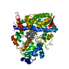
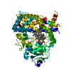
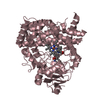
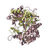
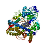
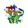
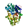
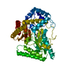
| Title | Crystal structure of Pif1 helicase from Bacteroides double mutant L95C-I339C | |||||||||
 Components Components | TPR DOMAIN PROTEIN | |||||||||
 Keywords Keywords | HYDROLASE / SF1B / G QUADRUPLEX / SH3 DOMAIN / CONFORMATIONAL CHANGE / DISULPHIDE BRIDGE | |||||||||
| Function / homology |  Function and homology information Function and homology informationtelomere maintenance / DNA helicase activity / nucleotide binding / DNA repair / metal ion binding Similarity search - Function | |||||||||
| Biological species |  BACTEROIDES (bacteria) BACTEROIDES (bacteria) | |||||||||
| Method |  X-RAY DIFFRACTION / SYNCHROTRON / MOLECULAR REPLACEMENT / Resolution: 2.412 Å X-RAY DIFFRACTION / SYNCHROTRON / MOLECULAR REPLACEMENT / Resolution: 2.412 Å | |||||||||
 Authors Authors | Chen, W.-F. / Dai, Y.-X. / Duan, X.-L. / Liu, N.-N. / Shi, W. / Li, M. / Dou, S.-X. / Li, N. / Dong, Y.-H. / Rety, S. / Xi, X.-G. | |||||||||
 Citation Citation | Journal: Nucleic Acids Res. / Year: 2016 Title: Crystal Structures of the Bspif1 Helicase Reveal that a Major Movement of the 2B SH3 Domain is Required for DNA Unwinding Authors: Chen, W.-F. / Dai, Y.-X. / Duan, X.-L. / Liu, N.-N. / Shi, W. / Li, N. / Li, M. / Dou, S.-X. / Dong, Y.-H. / Rety, S. / Xi, X.-G. | |||||||||
| History |
|
- Structure visualization
| Structure viewer | Molecule: MolmilJmol/JSmol |
|---|
- Downloads & links
-Download
| PDBx/mmCIF format | 5ftf.cif.gz | 173.6 KB | Display | PDBx/mmCIF format |
|---|---|---|---|---|
| PDB format | pdb5ftf.ent.gz | 139.4 KB | Display | PDB format |
| PDBx/mmJSON format | 5ftf.json.gz | Tree view | PDBx/mmJSON format | |
| Others |  Other downloads Other downloads |
-Validation report
| Arichive directory | https://data.pdbj.org/pub/pdb/validation_reports/ft/5ftfftp://data.pdbj.org/pub/pdb/validation_reports/ft/5ftf | HTTPS FTP |
|---|
-Related structure data
| Related structure data |  5ftbSC  5ftcC  5ftdC 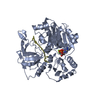 5fteC S: Starting model for refinement C: citing same article ( |
|---|---|
| Similar structure data |


-Links
 PDBj
PDBj
- Assembly
| Deposited unit | 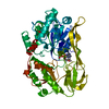
| ||||||||
|---|---|---|---|---|---|---|---|---|---|
| 1 |
| ||||||||
| Unit cell |
|
-Components
| #1: Protein | Mass: 49710.938 Da / Num. of mol.: 1 / Mutation: YES Source method: isolated from a genetically manipulated source Source: (gene. exp.) BACTEROIDES (bacteria) / Strain: SP. 3_1_23 / Production host: |
|---|---|
| #2: Chemical | ChemComp-ADP /   Mass: 427.201 Da / Num. of mol.: 1 / Source method: obtained synthetically / Formula: C10H15N5O10P2 / Comment: ADP, energy-carrying molecule*YM Mass: 427.201 Da / Num. of mol.: 1 / Source method: obtained synthetically / Formula: C10H15N5O10P2 / Comment: ADP, energy-carrying molecule*YM |
| #3: Water | ChemComp-HOH /  Mass: 18.015 Da / Num. of mol.: 30 / Source method: isolated from a natural source / Formula: H2O Mass: 18.015 Da / Num. of mol.: 30 / Source method: isolated from a natural source / Formula: H2O |
| Has protein modification | Y |
| Sequence details | NCBI REFERENCE SEQUENCE WP_008647876.1 |
-Experimental details
-Experiment
| Experiment | Method: X-RAY DIFFRACTION / Number of used crystals: 1 |
|---|
- Sample preparation
| Crystal | Density Matthews: 2.68 Å3/Da / Density % sol: 54.1 % / Description: NONE |
|---|---|
| Crystal grow | pH: 6.5 Details: 0.1M BIS-TRIS PROPANE PH 6.5 0.1M CALCIUM ACETATE 10% PEG 4000 |
-Data collection
| Diffraction | Mean temperature: 77 K |
|---|---|
| Diffraction source | Source: SYNCHROTRON / Site: SSRF  / Beamline: BL17U / Wavelength: 0.979142 / Beamline: BL17U / Wavelength: 0.979142 |
| Detector | Type: ADSC QUANTUM 315r / Detector: CCD / Date: Oct 13, 2015 |
| Radiation | Protocol: SINGLE WAVELENGTH / Monochromatic (M) / Laue (L): M / Scattering type: x-ray |
| Radiation wavelength | Wavelength: 0.979142 Å / Relative weight: 1 |
| Reflection | Resolution: 2.41→60.87 Å / Num. obs: 19410 / % possible obs: 90 % / Observed criterion σ(I): 2 / Redundancy: 7.1 % / Biso Wilson estimate: 52.73 Å2 / Rmerge(I) obs: 0.07 / Net I/σ(I): 16.15 |
| Reflection shell | Resolution: 2.41→2.5 Å / Redundancy: 7.4 % / Rmerge(I) obs: 0.81 / Mean I/σ(I) obs: 2.57 / % possible all: 97 |
- Processing
| Software |
| ||||||||||||||||||||||||||||||||||||||||||||||||||||||||
|---|---|---|---|---|---|---|---|---|---|---|---|---|---|---|---|---|---|---|---|---|---|---|---|---|---|---|---|---|---|---|---|---|---|---|---|---|---|---|---|---|---|---|---|---|---|---|---|---|---|---|---|---|---|---|---|---|---|
| Refinement | Method to determine structure: MOLECULAR REPLACEMENT Starting model: PDB ENTRY 5FTB Resolution: 2.412→60.868 Å / SU ML: 0.37 / σ(F): 1.34 / Phase error: 34.19 / Stereochemistry target values: ML
| ||||||||||||||||||||||||||||||||||||||||||||||||||||||||
| Solvent computation | Shrinkage radii: 0.9 Å / VDW probe radii: 1.11 Å / Solvent model: FLAT BULK SOLVENT MODEL | ||||||||||||||||||||||||||||||||||||||||||||||||||||||||
| Refinement step | Cycle: LAST / Resolution: 2.412→60.868 Å
| ||||||||||||||||||||||||||||||||||||||||||||||||||||||||
| Refine LS restraints |
| ||||||||||||||||||||||||||||||||||||||||||||||||||||||||
| LS refinement shell |
|
