 Movie
Movie Controller

[English] 日本語
 Yorodumi
- PDB-5jp1: Structure of Xanthomonas campestris effector protein XopD bound t... -
+ Open data

- Basic information
| Entry | Database: PDB / ID: 5jp1 | |||||||||
|---|---|---|---|---|---|---|---|---|---|---|
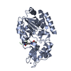
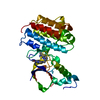
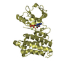
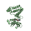
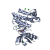
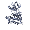
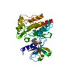
| Title | Structure of Xanthomonas campestris effector protein XopD bound to tomato SUMO | |||||||||
 Components Components |
| |||||||||
 Keywords Keywords | HYDROLASE / Enzyme / CE clan / Deubiquitinase / DeSUMOylase | |||||||||
| Function / homology |  Function and homology information Function and homology informationdeNEDDylase activity / protein deneddylation / ubiquitin-like protein ligase binding / protein sumoylation / cysteine-type peptidase activity / protein tag activity / proteolysis / nucleus Similarity search - Function | |||||||||
| Biological species |  Xanthomonas campestris pv. vesicatoria (bacteria) Xanthomonas campestris pv. vesicatoria (bacteria) | |||||||||
| Method |  X-RAY DIFFRACTION / SYNCHROTRON / MOLECULAR REPLACEMENT / Resolution: 2.1 Å X-RAY DIFFRACTION / SYNCHROTRON / MOLECULAR REPLACEMENT / Resolution: 2.1 Å | |||||||||
 Authors Authors | Pruneda, J.N. / Komander, D. | |||||||||
| Funding support |  United Kingdom, 1items United Kingdom, 1items
| |||||||||
 Citation Citation | Journal: Mol.Cell / Year: 2016 Title: The Molecular Basis for Ubiquitin and Ubiquitin-like Specificities in Bacterial Effector Proteases. Authors: Pruneda, J.N. / Durkin, C.H. / Geurink, P.P. / Ovaa, H. / Santhanam, B. / Holden, D.W. / Komander, D. | |||||||||
| History |
|
- Structure visualization
| Structure viewer | Molecule: MolmilJmol/JSmol |
|---|
- Downloads & links
-Download
| PDBx/mmCIF format | 5jp1.cif.gz | 129.4 KB | Display | PDBx/mmCIF format |
|---|---|---|---|---|
| PDB format | pdb5jp1.ent.gz | 98.7 KB | Display | PDB format |
| PDBx/mmJSON format | 5jp1.json.gz | Tree view | PDBx/mmJSON format | |
| Others |  Other downloads Other downloads |
-Validation report
| Summary document | 5jp1_validation.pdf.gz | 471.7 KB | Display | wwPDB validaton report |
|---|---|---|---|---|
| Full document | 5jp1_full_validation.pdf.gz | 489.7 KB | Display | |
| Data in XML | 5jp1_validation.xml.gz | 17.4 KB | Display | |
| Data in CIF | 5jp1_validation.cif.gz | 22.6 KB | Display | |
| Arichive directory | https://data.pdbj.org/pub/pdb/validation_reports/jp/5jp1ftp://data.pdbj.org/pub/pdb/validation_reports/jp/5jp1 | HTTPS FTP |
-Related structure data
| Related structure data |  5hafC 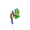 5hagC  5hamC  5jp3C 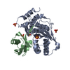 1tgzS  2oivS S: Starting model for refinement C: citing same article ( |
|---|---|
| Similar structure data |


-Links
 PDBj
PDBj- Assembly
| Deposited unit | 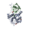
| |||||||||
|---|---|---|---|---|---|---|---|---|---|---|
| 1 |
| |||||||||
| Unit cell |
| |||||||||
| Components on special symmetry positions |
|
-Components
| #1: Protein | Mass: 24438.215 Da / Num. of mol.: 1 / Fragment: UNP residues 298-515 Source method: isolated from a genetically manipulated source Source: (gene. exp.) Xanthomonas campestris pv. vesicatoria (strain 85-10) (bacteria)Gene: xopD, XCV0437 / Production host: | ||||||
|---|---|---|---|---|---|---|---|
| #2: Protein | Mass: 10879.215 Da / Num. of mol.: 1 Source method: isolated from a genetically manipulated source Source: (gene. exp.) | ||||||
| #3: Chemical |   Mass: 102.046 Da / Num. of mol.: 2 / Source method: obtained synthetically / Formula: C3H2O4 Mass: 102.046 Da / Num. of mol.: 2 / Source method: obtained synthetically / Formula: C3H2O4#4: Chemical | ChemComp-D1D / ( |   Mass: 152.235 Da / Num. of mol.: 1 / Source method: obtained synthetically / Formula: C4H8O2S2 Mass: 152.235 Da / Num. of mol.: 1 / Source method: obtained synthetically / Formula: C4H8O2S2#5: Water | ChemComp-HOH / |  Mass: 18.015 Da / Num. of mol.: 199 / Source method: isolated from a natural source / Formula: H2O Mass: 18.015 Da / Num. of mol.: 199 / Source method: isolated from a natural source / Formula: H2OHas protein modification | Y | |
-Experimental details
-Experiment
| Experiment | Method: X-RAY DIFFRACTION / Number of used crystals: 1 |
|---|
- Sample preparation
| Crystal | Density Matthews: 2.93 Å3/Da / Density % sol: 58.02 % |
|---|---|
| Crystal grow | Temperature: 291 K / Method: vapor diffusion, sitting drop / Details: 0.1M bicine (pH 9.0) 1.6M ammonium sulfate |
-Data collection
| Diffraction | Mean temperature: 100 K |
|---|---|
| Diffraction source | Source: SYNCHROTRON / Site: Diamond / Beamline: I04-1 / Wavelength: 0.9282 Å |
| Detector | Type: DECTRIS PILATUS 6M / Detector: PIXEL / Date: Jan 16, 2016 |
| Radiation | Protocol: SINGLE WAVELENGTH / Monochromatic (M) / Laue (L): M / Scattering type: x-ray |
| Radiation wavelength | Wavelength: 0.9282 Å / Relative weight: 1 |
| Reflection | Resolution: 2.1→59.55 Å / Num. obs: 23605 / % possible obs: 98.1 % / Redundancy: 3.5 % / Rmerge(I) obs: 0.089 / Net I/σ(I): 8.7 |
| Reflection shell | Resolution: 2.1→2.16 Å / Redundancy: 3.4 % / Rmerge(I) obs: 0.465 / Mean I/σ(I) obs: 2 / % possible all: 99.4 |
- Processing
| Software |
| |||||||||||||||||||||||||||||||||||||||||||||||||||||||||||||||||||||||||||
|---|---|---|---|---|---|---|---|---|---|---|---|---|---|---|---|---|---|---|---|---|---|---|---|---|---|---|---|---|---|---|---|---|---|---|---|---|---|---|---|---|---|---|---|---|---|---|---|---|---|---|---|---|---|---|---|---|---|---|---|---|---|---|---|---|---|---|---|---|---|---|---|---|---|---|---|---|
| Refinement | Method to determine structure: MOLECULAR REPLACEMENT Starting model: 2OIV, 1TGZ Resolution: 2.1→59.551 Å / SU ML: 0.22 / Cross valid method: FREE R-VALUE / σ(F): 1.34 / Phase error: 20.12 / Stereochemistry target values: ML
| |||||||||||||||||||||||||||||||||||||||||||||||||||||||||||||||||||||||||||
| Solvent computation | Shrinkage radii: 0.9 Å / VDW probe radii: 1.11 Å / Solvent model: FLAT BULK SOLVENT MODEL | |||||||||||||||||||||||||||||||||||||||||||||||||||||||||||||||||||||||||||
| Refinement step | Cycle: LAST / Resolution: 2.1→59.551 Å
| |||||||||||||||||||||||||||||||||||||||||||||||||||||||||||||||||||||||||||
| Refine LS restraints |
| |||||||||||||||||||||||||||||||||||||||||||||||||||||||||||||||||||||||||||
| LS refinement shell |
| |||||||||||||||||||||||||||||||||||||||||||||||||||||||||||||||||||||||||||
| Refinement TLS params. | Method: refined / Refine-ID: X-RAY DIFFRACTION
| |||||||||||||||||||||||||||||||||||||||||||||||||||||||||||||||||||||||||||
| Refinement TLS group |
|
