| Entry | Database: PDB / ID: 5jg8
|
|---|
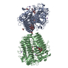
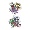
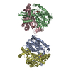
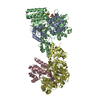
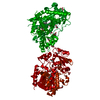
| Title | Crystal structure of human acid sphingomyelinase |
|---|
 Components Components | Sphingomyelin phosphodiesterase |
|---|
 Keywords Keywords | HYDROLASE / Lysosomal hydrolase / Neimann-Pick disease / Sphingolipid / Saposin |
|---|
| Function / homology |  Function and homology information Function and homology information
acid sphingomyelin phosphodiesterase activity / sphingomyelin metabolic process / sphingomyelin catabolic process / sphingomyelin phosphodiesterase / sphingomyelin phosphodiesterase activity / phospholipase C / phosphatidylcholine phospholipase C activity / endolysosome / lamellar body / termination of signal transduction ...acid sphingomyelin phosphodiesterase activity / sphingomyelin metabolic process / sphingomyelin catabolic process / sphingomyelin phosphodiesterase / sphingomyelin phosphodiesterase activity / phospholipase C / phosphatidylcholine phospholipase C activity / endolysosome / lamellar body / termination of signal transduction / glycosphingolipid catabolic process / Glycosphingolipid catabolism / plasma membrane repair / hydrolase activity, acting on glycosyl bonds / response to type I interferon / ceramide biosynthetic process / response to ionizing radiation / response to tumor necrosis factor / positive regulation of endocytosis / cholesterol metabolic process / negative regulation of MAPK cascade / lysosomal lumen / lipid droplet / cellular response to calcium ion / response to interleukin-1 / wound healing / response to cocaine / response to virus / cellular response to UV / nervous system development / positive regulation of viral entry into host cell / lysosome / endosome / response to xenobiotic stimulus / positive regulation of apoptotic process / symbiont entry into host cell / signal transduction / : / extracellular exosome / zinc ion binding / plasma membraneSimilarity search - Function Sphingomyelin phosphodiesterase / Acid sphingomyelinase/endopolyphosphatase, metallophosphatase domain / Sphingomyelin phosphodiesterase, C-terminal domain / Acid sphingomyelin phosphodiesterase C-terminal region / Saposin (B) Domains / Saposin B type domain / Saposin-like / Saposin B type domain profile. / Calcineurin-like phosphoesterase domain, ApaH type / Calcineurin-like phosphoesterase / Metallo-dependent phosphatase-likeSimilarity search - Domain/homology |
|---|
| Biological species |  Homo sapiens (human) Homo sapiens (human) |
|---|
| Method |  X-RAY DIFFRACTION / SYNCHROTRON / MOLECULAR REPLACEMENT / Resolution: 2.8 Å X-RAY DIFFRACTION / SYNCHROTRON / MOLECULAR REPLACEMENT / Resolution: 2.8 Å |
|---|
 Authors Authors | Xiong, Z.J. / Prive, G.G. |
|---|
 Citation Citation | Journal: J.Mol.Biol. / Year: 2016
Title: Structure of Human Acid Sphingomyelinase Reveals the Role of the Saposin Domain in Activating Substrate Hydrolysis.
Authors: Xiong, Z.J. / Huang, J. / Poda, G. / Pomes, R. / Prive, G.G. |
|---|
| History | | Deposition | Apr 19, 2016 | Deposition site: RCSB / Processing site: RCSB |
|---|
| Revision 1.0 | Jul 6, 2016 | Provider: repository / Type: Initial release |
|---|
| Revision 1.1 | Jul 13, 2016 | Group: Database references |
|---|
| Revision 1.2 | Aug 17, 2016 | Group: Database references |
|---|
| Revision 2.0 | Jul 29, 2020 | Group: Advisory / Atomic model ...Advisory / Atomic model / Data collection / Database references / Derived calculations / Structure summary
Category: atom_site / chem_comp ...atom_site / chem_comp / citation / entity / pdbx_branch_scheme / pdbx_chem_comp_identifier / pdbx_entity_branch / pdbx_entity_branch_descriptor / pdbx_entity_branch_link / pdbx_entity_branch_list / pdbx_entity_nonpoly / pdbx_nonpoly_scheme / pdbx_struct_assembly_gen / pdbx_struct_conn_angle / pdbx_struct_oper_list / pdbx_validate_close_contact / struct_asym / struct_conn / struct_site / struct_site_gen
Item: _atom_site.B_iso_or_equiv / _atom_site.Cartn_x ..._atom_site.B_iso_or_equiv / _atom_site.Cartn_x / _atom_site.Cartn_y / _atom_site.Cartn_z / _atom_site.auth_asym_id / _atom_site.auth_atom_id / _atom_site.auth_comp_id / _atom_site.auth_seq_id / _atom_site.label_asym_id / _atom_site.label_atom_id / _atom_site.label_comp_id / _atom_site.label_entity_id / _atom_site.type_symbol / _chem_comp.name / _chem_comp.type / _citation.journal_id_CSD / _pdbx_struct_assembly_gen.asym_id_list / _pdbx_struct_conn_angle.ptnr2_label_asym_id / _pdbx_struct_conn_angle.ptnr3_label_asym_id / _pdbx_struct_oper_list.symmetry_operation / _pdbx_validate_close_contact.auth_asym_id_1 / _pdbx_validate_close_contact.auth_asym_id_2 / _pdbx_validate_close_contact.auth_seq_id_1 / _pdbx_validate_close_contact.auth_seq_id_2 / _struct_conn.conn_type_id / _struct_conn.id / _struct_conn.pdbx_dist_value / _struct_conn.pdbx_leaving_atom_flag / _struct_conn.pdbx_role / _struct_conn.ptnr1_auth_asym_id / _struct_conn.ptnr1_auth_comp_id / _struct_conn.ptnr1_auth_seq_id / _struct_conn.ptnr1_label_asym_id / _struct_conn.ptnr1_label_atom_id / _struct_conn.ptnr1_label_comp_id / _struct_conn.ptnr1_label_seq_id / _struct_conn.ptnr2_auth_asym_id / _struct_conn.ptnr2_auth_comp_id / _struct_conn.ptnr2_auth_seq_id / _struct_conn.ptnr2_label_asym_id / _struct_conn.ptnr2_label_atom_id / _struct_conn.ptnr2_label_comp_id
Description: Carbohydrate remediation / Provider: repository / Type: Remediation |
|---|
| Revision 2.1 | Sep 27, 2023 | Group: Data collection / Database references ...Data collection / Database references / Refinement description / Structure summary
Category: chem_comp / chem_comp_atom ...chem_comp / chem_comp_atom / chem_comp_bond / database_2 / pdbx_initial_refinement_model / struct_ncs_dom_lim
Item: _chem_comp.pdbx_synonyms / _database_2.pdbx_DOI ..._chem_comp.pdbx_synonyms / _database_2.pdbx_DOI / _database_2.pdbx_database_accession / _struct_ncs_dom_lim.beg_auth_comp_id / _struct_ncs_dom_lim.beg_label_asym_id / _struct_ncs_dom_lim.beg_label_comp_id / _struct_ncs_dom_lim.beg_label_seq_id / _struct_ncs_dom_lim.end_auth_comp_id / _struct_ncs_dom_lim.end_label_asym_id / _struct_ncs_dom_lim.end_label_comp_id / _struct_ncs_dom_lim.end_label_seq_id |
|---|
| Revision 2.2 | Oct 23, 2024 | Group: Structure summary / Category: pdbx_entry_details / pdbx_modification_feature |
|---|
|
|---|
 Movie
Movie Controller


 Open data
 Basic information
 Structure visualization
 Downloads & links
 Other downloads


 PDBj
PDBj
 Assembly