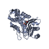
 Movie
Movie Controller


+ Open data

- Basic information
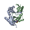
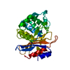
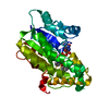
| Entry | Database: PDB / ID: 5jfz | ||||||
|---|---|---|---|---|---|---|---|
| Title | E. coli EcFicT in complex with EcFicA mutant E28G | ||||||
 Components Components |
| ||||||
 Keywords Keywords | TRANSFERASE / FIC domain / fic-1 / Class I Fic protein | ||||||
| Function / homology |  Function and homology information Function and homology informationAMPylase activity / protein adenylyltransferase / regulation of cell division / ATP binding Similarity search - Function | ||||||
| Biological species |  | ||||||
| Method |  X-RAY DIFFRACTION / SYNCHROTRON / MOLECULAR REPLACEMENT / molecular replacement / Resolution: 2.4 Å X-RAY DIFFRACTION / SYNCHROTRON / MOLECULAR REPLACEMENT / molecular replacement / Resolution: 2.4 Å | ||||||
 Authors Authors | Stanger, F.V. / Schirmer, T. | ||||||
 Citation Citation | Journal: Plos One / Year: 2016 Title: Crystal Structure of the Escherichia coli Fic Toxin-Like Protein in Complex with Its Cognate Antitoxin. Authors: Stanger, F.V. / Harms, A. / Dehio, C. / Schirmer, T. | ||||||
| History |
|
- Structure visualization
| Structure viewer | Molecule: MolmilJmol/JSmol |
|---|
- Downloads & links
-Download
| PDBx/mmCIF format | 5jfz.cif.gz | 149.2 KB | Display | PDBx/mmCIF format |
|---|---|---|---|---|
| PDB format | pdb5jfz.ent.gz | 116 KB | Display | PDB format |
| PDBx/mmJSON format | 5jfz.json.gz | Tree view | PDBx/mmJSON format | |
| Others |  Other downloads Other downloads |
-Validation report
| Arichive directory | https://data.pdbj.org/pub/pdb/validation_reports/jf/5jfzftp://data.pdbj.org/pub/pdb/validation_reports/jf/5jfz | HTTPS FTP |
|---|
-Related structure data
| Related structure data | 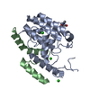 5jffC 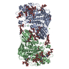 5fjjS S: Starting model for refinement C: citing same article ( |
|---|---|
| Similar structure data |


-Links
 PDBj
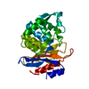
PDBj- Assembly
| Deposited unit | 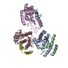
| ||||||||
|---|---|---|---|---|---|---|---|---|---|
| 1 | 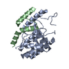
| ||||||||
| 2 | 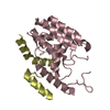
| ||||||||
| 3 | 
| ||||||||
| Unit cell |
|
-Components
| #1: Protein | Mass: 25627.748 Da / Num. of mol.: 3 Source method: isolated from a genetically manipulated source Source: (gene. exp.) References: UniProt: P20605, Transferases; Transferring phosphorus-containing groups; Nucleotidyltransferases #2: Protein | Mass: 7936.075 Da / Num. of mol.: 3 / Mutation: E28G Source method: isolated from a genetically manipulated source Source: (gene. exp.) #3: Water | ChemComp-HOH / |  Mass: 18.015 Da / Num. of mol.: 147 / Source method: isolated from a natural source / Formula: H2O Mass: 18.015 Da / Num. of mol.: 147 / Source method: isolated from a natural source / Formula: H2O |
|---|
-Experimental details
-Experiment
| Experiment | Method: X-RAY DIFFRACTION / Number of used crystals: 1 |
|---|
- Sample preparation
| Crystal | Density Matthews: 2.5 Å3/Da / Density % sol: 50.9 % / Mosaicity: 0.25 ° |
|---|---|
| Crystal grow | Temperature: 293.15 K / Method: vapor diffusion, sitting drop / pH: 6 Details: 19% PEG 1500 (w/v), 0.1 M MMT (malic acid, MES, Tris) buffer pH 6.0 |
-Data collection
| Diffraction | Mean temperature: 100 K | ||||||||||||||||||||||||||||||
|---|---|---|---|---|---|---|---|---|---|---|---|---|---|---|---|---|---|---|---|---|---|---|---|---|---|---|---|---|---|---|---|
| Diffraction source | Source: SYNCHROTRON / Site: SLS  / Beamline: X06SA / Wavelength: 1 Å / Beamline: X06SA / Wavelength: 1 Å | ||||||||||||||||||||||||||||||
| Detector | Type: DECTRIS PILATUS 6M / Detector: PIXEL / Date: Oct 31, 2012 | ||||||||||||||||||||||||||||||
| Radiation | Protocol: SINGLE WAVELENGTH / Monochromatic (M) / Laue (L): M / Scattering type: x-ray | ||||||||||||||||||||||||||||||
| Radiation wavelength | Wavelength: 1 Å / Relative weight: 1 | ||||||||||||||||||||||||||||||
| Reflection | Resolution: 2.4→80.41 Å / Num. obs: 30850 / % possible obs: 98.6 % / Redundancy: 6.2 % / CC1/2: 0.992 / Rmerge(I) obs: 0.176 / Rpim(I) all: 0.076 / Rrim(I) all: 0.193 / Net I/σ(I): 7.2 / Num. measured all: 192193 | ||||||||||||||||||||||||||||||
| Reflection shell | Diffraction-ID: 1 / Rejects: _
|
-Phasing
| Phasing | Method: molecular replacement | |||||||||
|---|---|---|---|---|---|---|---|---|---|---|
| Phasing MR | Model details: Phaser MODE: MR_AUTO
|
- Processing
| Software |
| |||||||||||||||||||||||||||||||||||||||||||||||||||||||||||||||||||||||||||
|---|---|---|---|---|---|---|---|---|---|---|---|---|---|---|---|---|---|---|---|---|---|---|---|---|---|---|---|---|---|---|---|---|---|---|---|---|---|---|---|---|---|---|---|---|---|---|---|---|---|---|---|---|---|---|---|---|---|---|---|---|---|---|---|---|---|---|---|---|---|---|---|---|---|---|---|---|
| Refinement | Method to determine structure: MOLECULAR REPLACEMENT Starting model: 5fjj Resolution: 2.4→80.41 Å / Cor.coef. Fo:Fc: 0.921 / Cor.coef. Fo:Fc free: 0.886 / SU B: 10.147 / SU ML: 0.234 / Cross valid method: THROUGHOUT / σ(F): 0 / ESU R: 0.484 / ESU R Free: 0.292 / Stereochemistry target values: MAXIMUM LIKELIHOOD Details: HYDROGENS HAVE BEEN ADDED IN THE RIDING POSITIONS U VALUES : REFINED INDIVIDUALLY
| |||||||||||||||||||||||||||||||||||||||||||||||||||||||||||||||||||||||||||
| Solvent computation | Ion probe radii: 0.8 Å / Shrinkage radii: 0.8 Å / VDW probe radii: 1.2 Å / Solvent model: MASK | |||||||||||||||||||||||||||||||||||||||||||||||||||||||||||||||||||||||||||
| Displacement parameters | Biso max: 104.93 Å2 / Biso mean: 32.801 Å2 / Biso min: 12.84 Å2
| |||||||||||||||||||||||||||||||||||||||||||||||||||||||||||||||||||||||||||
| Refinement step | Cycle: final / Resolution: 2.4→80.41 Å
| |||||||||||||||||||||||||||||||||||||||||||||||||||||||||||||||||||||||||||
| Refine LS restraints |
| |||||||||||||||||||||||||||||||||||||||||||||||||||||||||||||||||||||||||||
| LS refinement shell | Resolution: 2.398→2.46 Å / Total num. of bins used: 20
|
