 Movie
Movie Controller


+ Open data

- Basic information
| Entry | Database: PDB / ID: 1kgf | ||||||
|---|---|---|---|---|---|---|---|
| Title | STRUCTURE OF BETA-LACTAMASE ASN 170 GLN MUTANT | ||||||
 Components Components | BETA-LACTAMASE | ||||||
 Keywords Keywords | HYDROLASE / ANTIBIOTIC RESISTANCE / PLASMID / TRANSPOSABLE ELEMENT | ||||||
| Function / homology |  Function and homology information Function and homology informationbeta-lactam antibiotic catabolic process / beta-lactamase activity / beta-lactamase / response to antibiotic Similarity search - Function | ||||||
| Biological species |   Staphylococcus aureus (bacteria) Staphylococcus aureus (bacteria) | ||||||
| Method |  X-RAY DIFFRACTION / Resolution: 2.2 Å X-RAY DIFFRACTION / Resolution: 2.2 Å | ||||||
 Authors Authors | Chen, C.C.H. / Zawadzke, L.E. / Herzberg, O. | ||||||
 Citation Citation | Journal: Biochemistry / Year: 1996 Title: Elimination of the hydrolytic water molecule in a class A beta-lactamase mutant: crystal structure and kinetics. Authors: Zawadzke, L.E. / Chen, C.C. / Banerjee, S. / Li, Z. / Wasch, S. / Kapadia, G. / Moult, J. / Herzberg, O. #1: Journal: Biochemistry / Year: 1996Title: Structure and Kinetics of the Beta-Lactamase Mutants S70A and K73H from Staphylococcus Aureus Pc1 Authors: Chen, C.C. / Smith, T.J. / Kapadia, G. / Wasch, S. / Zawadzke, L.E. / Coulson, A. / Herzberg, O. #2: Journal: Protein Eng. / Year: 1995Title: An Engineered Staphylococcus Aureus Pc1 Beta-Lactamase that Hydrolyses Third-Generation Cephalosporins Authors: Zawadzke, L.E. / Smith, T.J. / Herzberg, O. #3: Journal: J.Mol.Biol. / Year: 1991Title: Refined Crystal Structure of Beta-Lactamase from Staphylococcus Aureus Pc1 at 2.0 Authors: Herzberg, O. #4: Journal: Science / Year: 1987Title: Bacterial Resistance to Beta-Lactam Antibiotics. Crystal Structure of Beta-Lactamase from Staphylococcus Aureus Pc1 at 2.5 A Resolution Authors: Herzberg, O. / Moult, J. | ||||||
| History |
| ||||||
| Remark 650 | HELIX SECONDARY STRUCTURE ASSIGNMENT IS ACCORDING TO W. KABSCH AND C. SANDER (BIOPOLYMERS 22, 2577- ...HELIX SECONDARY STRUCTURE ASSIGNMENT IS ACCORDING TO W. KABSCH AND C. SANDER (BIOPOLYMERS 22, 2577-2637, 1983). HELIX NUMBERING IS AS IN 3BLM. |
- Structure visualization
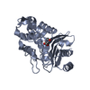
| Structure viewer | Molecule: MolmilJmol/JSmol |
|---|
- Downloads & links
-Download
| PDBx/mmCIF format | 1kgf.cif.gz | 67.6 KB | Display | PDBx/mmCIF format |
|---|---|---|---|---|
| PDB format | pdb1kgf.ent.gz | 49 KB | Display | PDB format |
| PDBx/mmJSON format | 1kgf.json.gz | Tree view | PDBx/mmJSON format | |
| Others |  Other downloads Other downloads |
-Validation report
| Arichive directory | https://data.pdbj.org/pub/pdb/validation_reports/kg/1kgfftp://data.pdbj.org/pub/pdb/validation_reports/kg/1kgf | HTTPS FTP |
|---|
-Related structure data


-Links
 PDBj
PDBj


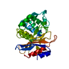
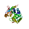
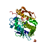
- Assembly
| Deposited unit | 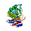
| ||||||||
|---|---|---|---|---|---|---|---|---|---|
| 1 |
| ||||||||
| Unit cell |
|
-Components
| #1: Protein | Mass: 28988.434 Da / Num. of mol.: 1 / Mutation: N170Q Source method: isolated from a genetically manipulated source Source: (gene. exp.) Staphylococcus aureus (bacteria) / Strain: PC1 / Description: INDUCIBLE TRC PROMOTER / Gene: BLAZ / Plasmid: PTS32 / Gene (production host): BLAZ / Production host: |
|---|---|
| #2: Water | ChemComp-HOH /  Mass: 18.015 Da / Num. of mol.: 219 / Source method: isolated from a natural source / Formula: H2O Mass: 18.015 Da / Num. of mol.: 219 / Source method: isolated from a natural source / Formula: H2O |
| Sequence details | RESIDUE NUMBERING IS BASED ON AMBLER, 1979 (R. P. AMBLER, BETA-LACTAMASES, J. M. T. HAMILTON-MILLER ...RESIDUE NUMBERING IS BASED ON AMBLER, 1979 (R. P. AMBLER, BETA-LACTAMASES |
-Experimental details
-Experiment
| Experiment | Method: X-RAY DIFFRACTION |
|---|
- Sample preparation
| Crystal | Density Matthews: 3.12 Å3/Da / Density % sol: 65 % | ||||||||||||||||||||||||||||||||||||||||||||||||||||||||||||
|---|---|---|---|---|---|---|---|---|---|---|---|---|---|---|---|---|---|---|---|---|---|---|---|---|---|---|---|---|---|---|---|---|---|---|---|---|---|---|---|---|---|---|---|---|---|---|---|---|---|---|---|---|---|---|---|---|---|---|---|---|---|
| Crystal grow | *PLUS pH: 8 / Method: vapor diffusion, hanging drop | ||||||||||||||||||||||||||||||||||||||||||||||||||||||||||||
| Components of the solutions | *PLUS
|
-Data collection
| Diffraction | Mean temperature: 293 K |
|---|---|
| Diffraction source | Wavelength: 1.5418 |
| Detector | Type: SIEMENS / Detector: AREA DETECTOR / Date: Dec 14, 1994 |
| Radiation | Monochromatic (M) / Laue (L): M / Scattering type: x-ray |
| Radiation wavelength | Wavelength: 1.5418 Å / Relative weight: 1 |
| Reflection | Num. obs: 16464 / % possible obs: 87 % / Observed criterion σ(I): 0 / Redundancy: 1.7 % / Rmerge(I) obs: 0.079 |
| Reflection | *PLUS Highest resolution: 2.2 Å / Num. measured all: 28166 |
| Reflection shell | *PLUS Highest resolution: 2.2 Å / Lowest resolution: 2.3 Å / % possible obs: 76 % / Rmerge(I) obs: 0.215 |
- Processing
| Software |
| ||||||||||||||||||||||||||||||||||||||||||||||||||||||||||||
|---|---|---|---|---|---|---|---|---|---|---|---|---|---|---|---|---|---|---|---|---|---|---|---|---|---|---|---|---|---|---|---|---|---|---|---|---|---|---|---|---|---|---|---|---|---|---|---|---|---|---|---|---|---|---|---|---|---|---|---|---|---|
| Refinement | Resolution: 2.2→8 Å / σ(F): 2 /
| ||||||||||||||||||||||||||||||||||||||||||||||||||||||||||||
| Refinement step | Cycle: LAST / Resolution: 2.2→8 Å
| ||||||||||||||||||||||||||||||||||||||||||||||||||||||||||||
| Refine LS restraints |
| ||||||||||||||||||||||||||||||||||||||||||||||||||||||||||||
| Software | *PLUS Name: X-PLOR / Classification: refinement | ||||||||||||||||||||||||||||||||||||||||||||||||||||||||||||
| Refinement | *PLUS | ||||||||||||||||||||||||||||||||||||||||||||||||||||||||||||
| Solvent computation | *PLUS | ||||||||||||||||||||||||||||||||||||||||||||||||||||||||||||
| Displacement parameters | *PLUS |
