 Movie
Movie Controller

[English] 日本語
 Yorodumi
- PDB-2a4x: Crystal Structure Of Mitomycin C-Binding Protein Complexed with M... -
+ Open data

- Basic information
| Entry | Database: PDB / ID: 2a4x | ||||||
|---|---|---|---|---|---|---|---|
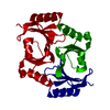
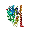
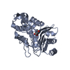
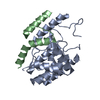
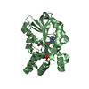
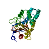
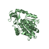
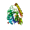
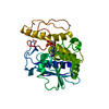
| Title | Crystal Structure Of Mitomycin C-Binding Protein Complexed with Metal-Free Bleomycin A2 | ||||||
 Components Components | Mitomycin-Binding Protein | ||||||
 Keywords Keywords | ANTIMICROBIAL PROTEIN / alfa/beta protein / Mitomycin C-binding protein / Bleomycin A2 / Structural Genomics | ||||||
| Function / homology |  Function and homology information Function and homology information2,3-Dihydroxybiphenyl 1,2-Dioxygenase, domain 1 / 2,3-Dihydroxybiphenyl 1,2-Dioxygenase; domain 1 / Glyoxalase/fosfomycin resistance/dioxygenase domain / Glyoxalase/Bleomycin resistance protein/Dioxygenase superfamily / Vicinal oxygen chelate (VOC) domain / Vicinal oxygen chelate (VOC) domain profile. / Glyoxalase/Bleomycin resistance protein/Dihydroxybiphenyl dioxygenase / Roll / Alpha Beta Similarity search - Domain/homology | ||||||
| Biological species |  Streptomyces caespitosus (bacteria) Streptomyces caespitosus (bacteria) | ||||||
| Method |  X-RAY DIFFRACTION / SYNCHROTRON / MOLECULAR REPLACEMENT / Resolution: 1.4 Å X-RAY DIFFRACTION / SYNCHROTRON / MOLECULAR REPLACEMENT / Resolution: 1.4 Å | ||||||
 Authors Authors | Danshiitsoodol, N. / de Pinho, C.A. / Matoba, Y. / Kumagai, T. / Sugiyama, M. | ||||||
 Citation Citation | Journal: J.Mol.Biol. / Year: 2006 Title: The Mitomycin C (MMC)-binding Protein from MMC-producing Microorganisms Protects from the Lethal Effect of Bleomycin: Crystallographic Analysis to Elucidate the Binding Mode of the Antibiotic to the Protein Authors: Danshiitsoodol, N. / de Pinho, C.A. / Matoba, Y. / Kumagai, T. / Sugiyama, M. | ||||||
| History |
|
- Structure visualization
| Structure viewer | Molecule: MolmilJmol/JSmol |
|---|
- Downloads & links
-Download
| PDBx/mmCIF format | 2a4x.cif.gz | 72.6 KB | Display | PDBx/mmCIF format |
|---|---|---|---|---|
| PDB format | pdb2a4x.ent.gz | 52 KB | Display | PDB format |
| PDBx/mmJSON format | 2a4x.json.gz | Tree view | PDBx/mmJSON format | |
| Others |  Other downloads Other downloads |
-Validation report
| Arichive directory | https://data.pdbj.org/pub/pdb/validation_reports/a4/2a4xftp://data.pdbj.org/pub/pdb/validation_reports/a4/2a4x | HTTPS FTP |
|---|
-Related structure data


-Links
 PDBj
PDBj- Assembly
| Deposited unit | 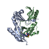
| ||||||||
|---|---|---|---|---|---|---|---|---|---|
| 1 |
| ||||||||
| Unit cell |
|
-Components
| #1: Protein | Mass: 15466.281 Da / Num. of mol.: 2 Source method: isolated from a genetically manipulated source Source: (gene. exp.) Streptomyces caespitosus (bacteria) / Gene: mrd / Plasmid: pET-mrd / Production host: #2: Chemical | ChemComp-BLM / |   Mass: 1416.560 Da / Num. of mol.: 1 / Source method: obtained synthetically / Formula: C55H85N17O21S3 / Comment: medication*YM Mass: 1416.560 Da / Num. of mol.: 1 / Source method: obtained synthetically / Formula: C55H85N17O21S3 / Comment: medication*YM#3: Water | ChemComp-HOH / |  Mass: 18.015 Da / Num. of mol.: 293 / Source method: isolated from a natural source / Formula: H2O Mass: 18.015 Da / Num. of mol.: 293 / Source method: isolated from a natural source / Formula: H2OSequence details | A SEQUENCE DATABASE REFERENCE FOR THIS PROTEIN WHICH DERIVES FROM STREPTOMYCES CAESPITOSUS DOES NOT ...A SEQUENCE DATABASE REFERENCE FOR THIS PROTEIN WHICH DERIVES FROM STREPTOMYC | |
|---|
-Experimental details
-Experiment
| Experiment | Method: X-RAY DIFFRACTION / Number of used crystals: 1 |
|---|
- Sample preparation
| Crystal | Density Matthews: 2.16 Å3/Da / Density % sol: 43.17 % |
|---|---|
| Crystal grow | Temperature: 298 K / Method: vapor diffusion, sitting drop / pH: 6 Details: PEG8000, MES, pH 6.0, VAPOR DIFFUSION, SITTING DROP, temperature 298K |
-Data collection
| Diffraction | Mean temperature: 100 K |
|---|---|
| Diffraction source | Source: SYNCHROTRON / Site: SPring-8  / Beamline: BL38B1 / Wavelength: 0.9 Å / Beamline: BL38B1 / Wavelength: 0.9 Å |
| Detector | Type: MARRESEARCH / Detector: CCD / Date: Feb 17, 2003 |
| Radiation | Protocol: SINGLE WAVELENGTH / Monochromatic (M) / Laue (L): M / Scattering type: x-ray |
| Radiation wavelength | Wavelength: 0.9 Å / Relative weight: 1 |
| Reflection | Resolution: 1.37→200 Å / Num. all: 57018 / Num. obs: 52971 / % possible obs: 91.3 % / Observed criterion σ(F): 1 / Observed criterion σ(I): 1 / Redundancy: 16 % / Biso Wilson estimate: 15.5 Å2 / Rmerge(I) obs: 0.041 / Net I/σ(I): 52.7 |
| Reflection shell | Resolution: 1.37→1.42 Å / Rmerge(I) obs: 0.481 / Mean I/σ(I) obs: 4.1 / Num. unique all: 4107 / % possible all: 72 |
- Processing
| Software |
| ||||||||||||||||||||||||||||||||||||
|---|---|---|---|---|---|---|---|---|---|---|---|---|---|---|---|---|---|---|---|---|---|---|---|---|---|---|---|---|---|---|---|---|---|---|---|---|---|
| Refinement | Method to determine structure: MOLECULAR REPLACEMENT / Resolution: 1.4→10 Å / Rfactor Rfree error: 0.005 / Data cutoff high absF: 10000000 / Data cutoff low absF: 0.001 / Isotropic thermal model: RESTRAINED / Cross valid method: THROUGHOUT / σ(F): 2 / Stereochemistry target values: Engh & Huber
| ||||||||||||||||||||||||||||||||||||
| Displacement parameters | Biso mean: 17.9 Å2
| ||||||||||||||||||||||||||||||||||||
| Refine analyze |
| ||||||||||||||||||||||||||||||||||||
| Refinement step | Cycle: LAST / Resolution: 1.4→10 Å
| ||||||||||||||||||||||||||||||||||||
| Refine LS restraints |
| ||||||||||||||||||||||||||||||||||||
| LS refinement shell | Resolution: 1.4→1.49 Å / Rfactor Rfree error: 0.017 / Total num. of bins used: 6
| ||||||||||||||||||||||||||||||||||||
| Xplor file |
|
