#191 - Nov 2015 Glutamate-gated Chloride Receptors similarity (4)
-
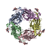
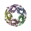
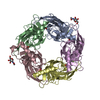
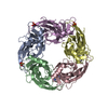
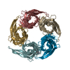
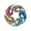
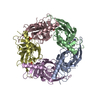
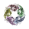
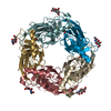
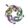
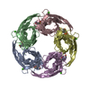
Assembly
Deposited unit
A: Acetylcholine-binding protein B: Acetylcholine-binding protein C: Acetylcholine-binding protein D: Acetylcholine-binding protein E: Acetylcholine-binding protein F: Acetylcholine-binding protein G: Acetylcholine-binding protein H: Acetylcholine-binding protein I: Acetylcholine-binding protein J: Acetylcholine-binding protein hetero molecules
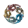
A: Acetylcholine-binding protein B: Acetylcholine-binding protein C: Acetylcholine-binding protein D: Acetylcholine-binding protein E: Acetylcholine-binding protein hetero molecules
F: Acetylcholine-binding protein G: Acetylcholine-binding protein H: Acetylcholine-binding protein I: Acetylcholine-binding protein J: Acetylcholine-binding protein hetero molecules
Monochromator: Si(220) Asymmetric cut single crystal / Protocol: SINGLE WAVELENGTH / Monochromatic (M) / Laue (L): M / Scattering type: x-ray
Radiation wavelength
Wavelength: 1 Å / Relative weight: 1
Reflection
Limit h max: 44 / Limit h min: -46 / Limit k max: 56 / Limit k min: 0 / Limit l max: 46 / Limit l min: 0 / Number: 120631 / D res high: 2.326 Å / D res low: 104.83 Å / Num. obs: 118832
In the structure databanks used in Yorodumi, some data are registered as the other names, "COVID-19 virus" and "2019-nCoV". Here are the details of the virus and the list of structure data.
Jan 31, 2019. EMDB accession codes are about to change! (news from PDBe EMDB page)
EMDB accession codes are about to change! (news from PDBe EMDB page)
The allocation of 4 digits for EMDB accession codes will soon come to an end. Whilst these codes will remain in use, new EMDB accession codes will include an additional digit and will expand incrementally as the available range of codes is exhausted. The current 4-digit format prefixed with “EMD-” (i.e. EMD-XXXX) will advance to a 5-digit format (i.e. EMD-XXXXX), and so on. It is currently estimated that the 4-digit codes will be depleted around Spring 2019, at which point the 5-digit format will come into force.
The EM Navigator/Yorodumi systems omit the EMD- prefix.
Related info.:Q: What is EMD? / ID/Accession-code notation in Yorodumi/EM Navigator
Yorodumi is a browser for structure data from EMDB, PDB, SASBDB, etc.
This page is also the successor to EM Navigator detail page, and also detail information page/front-end page for Omokage search.
The word "yorodu" (or yorozu) is an old Japanese word meaning "ten thousand". "mi" (miru) is to see.
Related info.:EMDB / PDB / SASBDB / Comparison of 3 databanks / Yorodumi Search / Aug 31, 2016. New EM Navigator & Yorodumi / Yorodumi Papers / Jmol/JSmol / Function and homology information / Changes in new EM Navigator and Yorodumi
 Movie
Movie Controller

 Yorodumi
 Open data
 Basic information
 Components
 Keywords
 Function and homology information
 Lymnaea stagnalis (great pond snail)
 X-RAY DIFFRACTION /
  Authors
 United States, 1items
  Citation
 Structure visualization
 Downloads & links
 Other downloads
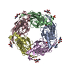


 PDBj
PDBj

 Assembly
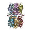

 Homo sapiens (human) / References: UniProt: P58154
 Mass: 398.460 Da / Num. of mol.: 10 / Source method: obtained synthetically / Formula: C23H22N6O
 Type: D-saccharide, beta linking / Mass: 221.208 Da / Num. of mol.: 10
 Mass: 94.971 Da / Num. of mol.: 11 / Source method: obtained synthetically / Formula: PO4
 Mass: 18.015 Da / Num. of mol.: 140 / Source method: isolated from a natural source / Formula: H2O
 Sample preparation
 Processing