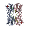
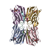
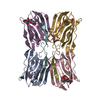
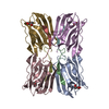
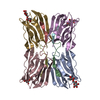
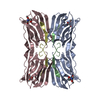
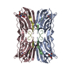
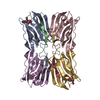
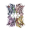
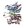
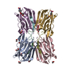
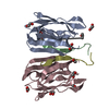
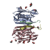
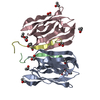
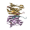
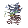
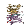
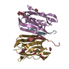
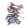
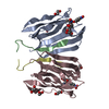
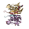
- PDB-5j50: Structure of tetrameric jacalin complexed with Gal beta-(1,3) Gal... -
+
Open data
ID or keywords:
Loading...
-
Basic information
Entry
Database: PDB / ID: 5j50
Title
Structure of tetrameric jacalin complexed with Gal beta-(1,3) GalNAc-alpha-OPNP
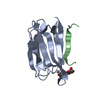
Components
Agglutinin alpha chain
Agglutinin beta-3 chain
Keywords
SUGAR BINDING PROTEIN / Plant lectins / Galactose specific lectin / beta-prism I fold / post translational proteolysis / T-antigen binding protein / reducing and non-reducing sugars
Resolution: 2.05→51.27 Å / Cor.coef. Fo:Fc: 0.927 / Cor.coef. Fo:Fc free: 0.887 / SU B: 10.405 / SU ML: 0.129 / Cross valid method: THROUGHOUT / ESU R: 0.229 / ESU R Free: 0.189 / Details: HYDROGENS HAVE NOT BEEN USED IN THE REFINEMENT
Rfactor
Num. reflection
% reflection
Selection details
Rfree
0.233
3892
10 %
RANDOM
Rwork
0.182
-
-
-
obs
0.187
35165
99.7 %
-
Solvent computation
Ion probe radii: 0.8 Å / Shrinkage radii: 0.8 Å / VDW probe radii: 1.2 Å
 Movie
Movie Controller

 Yorodumi
 Open data
 Basic information
 Components
 Keywords
 Function and homology information
 Artocarpus integer (chempedak)
 X-RAY DIFFRACTION /
  Authors
 India, 1items
  Citation
 Structure visualization
 Downloads & links
 Other downloads
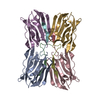


 PDBj
PDBj
 Assembly
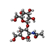


 Mass: 62.068 Da / Num. of mol.: 12 / Source method: obtained synthetically / Formula: C2H6O2
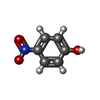
 Mass: 139.109 Da / Num. of mol.: 4 / Source method: obtained synthetically / Formula: C6H5NO3
 Mass: 106.120 Da / Num. of mol.: 1 / Source method: obtained synthetically / Formula: C4H10O3
 Sample preparation
 Processing