 Movie
Movie Controller


+ Open data

- Basic information
| Entry | Database: PDB / ID: 1ugw | ||||||
|---|---|---|---|---|---|---|---|
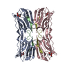
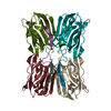
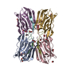
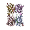
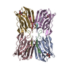
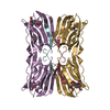
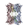
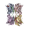
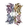
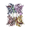
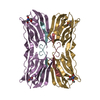
| Title | Crystal structure of jacalin- Gal complex | ||||||
 Components Components |
| ||||||
 Keywords Keywords | SUGAR BINDING PROTEIN / All beta sheet protein / Beta prism I fold / Galactose specific | ||||||
| Function / homology |  Function and homology information Function and homology information | ||||||
| Biological species |   Artocarpus integer (chempedak) Artocarpus integer (chempedak) | ||||||
| Method |  X-RAY DIFFRACTION / SYNCHROTRON / MOLECULAR REPLACEMENT / Resolution: 1.7 Å X-RAY DIFFRACTION / SYNCHROTRON / MOLECULAR REPLACEMENT / Resolution: 1.7 Å | ||||||
 Authors Authors | Jeyaprakash, A.A. / Katiyar, S. / Swaminathan, C.P. / Sekar, K. / Surolia, A. / Vijayan, M. | ||||||
 Citation Citation | Journal: J.MOL.BIOL. / Year: 2003 Title: Structural Basis of the Carbohydrate Specificities of Jacalin: An X-ray and Modeling Study Authors: Jeyaprakash, A.A. / Katiyar, S. / Swaminathan, C.P. / Sekar, K. / Surolia, A. / Vijayan, M. #1: Journal: J.Mol.Biol. / Year: 2002Title: Crystal structure of the jacalin-T-antigen complex and a comparative study of lectin-T-antigen complexes Authors: Jeyaprakash, A.A. / Rani, P.G. / Reddy, G.B. / Banumathi, S. / Betzel, C. / Sekar, K. / Surolia, A. / Vijayan, M. #2: Journal: Nat.Struct.Biol. / Year: 1996Title: A Novel mode of carbohydrate recognition in jacalin, A moraceae plant lectin with a beta-prism fold Authors: Sankaranarayanan, R. / Sekar, K. / Banerjee, R. / Sharma, V. / Surolia, A. / Vijayan, M. | ||||||
| History |
|
- Structure visualization
| Structure viewer | Molecule: MolmilJmol/JSmol |
|---|
- Downloads & links
-Download
| PDBx/mmCIF format | 1ugw.cif.gz | 136.5 KB | Display | PDBx/mmCIF format |
|---|---|---|---|---|
| PDB format | pdb1ugw.ent.gz | 106.8 KB | Display | PDB format |
| PDBx/mmJSON format | 1ugw.json.gz | Tree view | PDBx/mmJSON format | |
| Others |  Other downloads Other downloads |
-Validation report
| Arichive directory | https://data.pdbj.org/pub/pdb/validation_reports/ug/1ugwftp://data.pdbj.org/pub/pdb/validation_reports/ug/1ugw | HTTPS FTP |
|---|
-Related structure data
| Related structure data | 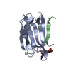 1ugxC  1ugyC  1uh0C 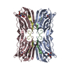 1uh1C  1m26S S: Starting model for refinement C: citing same article ( |
|---|---|
| Similar structure data |


-Links
 PDBj
PDBj

- Assembly
| Deposited unit | 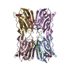
| ||||||||
|---|---|---|---|---|---|---|---|---|---|
| 1 |
| ||||||||
| Unit cell |
|
-Components
| #1: Protein | Mass: 14643.431 Da / Num. of mol.: 2 / Source method: isolated from a natural source / Source: (natural) Artocarpus integer (chempedak) / Organ: seeds / References: UniProt: P18670#2: Protein/peptide | Mass: 2047.204 Da / Num. of mol.: 4 / Source method: isolated from a natural source / Source: (natural) Artocarpus integer (chempedak) / Organ: seeds / References: UniProt: P18673#3: Protein | Mass: 14673.479 Da / Num. of mol.: 2 / Source method: isolated from a natural source / Source: (natural) Artocarpus integer (chempedak) / Organ: seeds / References: UniProt: P18670#4: Sugar | ChemComp-GAL /   Type: D-saccharide, beta linking / Mass: 180.156 Da / Num. of mol.: 4 Type: D-saccharide, beta linking / Mass: 180.156 Da / Num. of mol.: 4Source method: isolated from a genetically manipulated source Formula: C6H12O6 #5: Water | ChemComp-HOH / |  Mass: 18.015 Da / Num. of mol.: 489 / Source method: isolated from a natural source / Formula: H2O Mass: 18.015 Da / Num. of mol.: 489 / Source method: isolated from a natural source / Formula: H2O |
|---|
-Experimental details
-Experiment
| Experiment | Method: X-RAY DIFFRACTION / Number of used crystals: 1 |
|---|
- Sample preparation
| Crystal | Density Matthews: 2.94 Å3/Da / Density % sol: 57.86 % | |||||||||||||||||||||||||||||||||||||||||||||||||
|---|---|---|---|---|---|---|---|---|---|---|---|---|---|---|---|---|---|---|---|---|---|---|---|---|---|---|---|---|---|---|---|---|---|---|---|---|---|---|---|---|---|---|---|---|---|---|---|---|---|---|
| Crystal grow | Temperature: 293 K / Method: vapor diffusion, hanging drop / pH: 7.3 Details: PEG 4000, ammonium sulfate, sodium acetate trihydrate, pH 7.3, VAPOR DIFFUSION, HANGING DROP, temperature 293K | |||||||||||||||||||||||||||||||||||||||||||||||||
| Crystal grow | *PLUS Method: vapor diffusion | |||||||||||||||||||||||||||||||||||||||||||||||||
| Components of the solutions | *PLUS
|
-Data collection
| Diffraction | Mean temperature: 293 K |
|---|---|
| Diffraction source | Source: SYNCHROTRON / Site: ELETTRA  / Beamline: 5.2R / Wavelength: 1 Å / Beamline: 5.2R / Wavelength: 1 Å |
| Detector | Type: MARRESEARCH / Detector: IMAGE PLATE |
| Radiation | Monochromator: Mirrors / Protocol: SINGLE WAVELENGTH / Monochromatic (M) / Laue (L): M / Scattering type: x-ray |
| Radiation wavelength | Wavelength: 1 Å / Relative weight: 1 |
| Reflection | Resolution: 1.7→20 Å / Num. all: 91056 / Num. obs: 91056 / % possible obs: 96.8 % / Observed criterion σ(F): 0 / Observed criterion σ(I): 0 / Redundancy: 3.9 % / Biso Wilson estimate: 17.3 Å2 / Rmerge(I) obs: 0.055 |
| Reflection shell | Resolution: 1.7→1.76 Å / Rmerge(I) obs: 0.447 / Num. unique all: 8911 / % possible all: 95.8 |
| Reflection | *PLUS Highest resolution: 1.7 Å / Lowest resolution: 20 Å / Num. measured all: 350856 |
| Reflection shell | *PLUS % possible obs: 95.8 % / Num. unique obs: 8911 |
- Processing
| Software |
| ||||||||||||||||||||||||||||||||||||
|---|---|---|---|---|---|---|---|---|---|---|---|---|---|---|---|---|---|---|---|---|---|---|---|---|---|---|---|---|---|---|---|---|---|---|---|---|---|
| Refinement | Method to determine structure: MOLECULAR REPLACEMENT Starting model: 1M26 Resolution: 1.7→19.72 Å / Rfactor Rfree error: 0.003 / Data cutoff high absF: 2109195.61 / Data cutoff low absF: 0 / Isotropic thermal model: RESTRAINED / Cross valid method: THROUGHOUT / σ(F): 0 / Stereochemistry target values: Engh & Huber
| ||||||||||||||||||||||||||||||||||||
| Solvent computation | Solvent model: FLAT MODEL / Bsol: 49.9882 Å2 / ksol: 0.324271 e/Å3 | ||||||||||||||||||||||||||||||||||||
| Displacement parameters | Biso mean: 25.3 Å2
| ||||||||||||||||||||||||||||||||||||
| Refine analyze |
| ||||||||||||||||||||||||||||||||||||
| Refinement step | Cycle: LAST / Resolution: 1.7→19.72 Å
| ||||||||||||||||||||||||||||||||||||
| Refine LS restraints |
| ||||||||||||||||||||||||||||||||||||
| LS refinement shell | Resolution: 1.7→1.81 Å / Rfactor Rfree error: 0.011 / Total num. of bins used: 6
| ||||||||||||||||||||||||||||||||||||
| Xplor file |
| ||||||||||||||||||||||||||||||||||||
| Refinement | *PLUS Lowest resolution: 20 Å | ||||||||||||||||||||||||||||||||||||
| Solvent computation | *PLUS | ||||||||||||||||||||||||||||||||||||
| Displacement parameters | *PLUS | ||||||||||||||||||||||||||||||||||||
| Refine LS restraints | *PLUS
|
