 Movie
Movie Controller

[English] 日本語
 Yorodumi
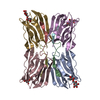
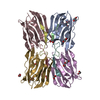
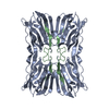
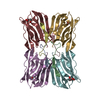
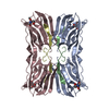
- PDB-1kuj: Crystal structure of Jacalin complexed with 1-O-methyl-alpha-D-mannose -
+ Open data

- Basic information
| Entry | Database: PDB / ID: 1kuj | ||||||
|---|---|---|---|---|---|---|---|
| Title | Crystal structure of Jacalin complexed with 1-O-methyl-alpha-D-mannose | ||||||
 Components Components |
| ||||||
 Keywords Keywords | SUGAR BINDING PROTEIN / LECTIN / BETA-PRISM FOLD / CARBOHYDRATE BINDING | ||||||
| Function / homology |  Function and homology information Function and homology information | ||||||
| Biological species |   Artocarpus integer (chempedak) Artocarpus integer (chempedak) | ||||||
| Method |  X-RAY DIFFRACTION / SYNCHROTRON / FOURIER SYNTHESIS / Resolution: 2 Å X-RAY DIFFRACTION / SYNCHROTRON / FOURIER SYNTHESIS / Resolution: 2 Å | ||||||
 Authors Authors | Bourne, Y. / Astoul, C.H. / Zamboni, V. / Peumans, W.J. / Menu-Bouaouiche, L. / Van Damme, E.J.M. / Barre, A. / Rouge, P. | ||||||
 Citation Citation | Journal: Biochem.J. / Year: 2002 Title: Structural basis for the unusual carbohydrate-binding specificity of jacalin towards galactose and mannose. Authors: Bourne, Y. / Astoul, C.H. / Zamboni, V. / Peumans, W.J. / Menu-Bouaouiche, L. / Van Damme, E.J. / Barre, A. / Rouge, P. | ||||||
| History |
|
- Structure visualization
| Structure viewer | Molecule: MolmilJmol/JSmol |
|---|
- Downloads & links
-Download
| PDBx/mmCIF format | 1kuj.cif.gz | 130.4 KB | Display | PDBx/mmCIF format |
|---|---|---|---|---|
| PDB format | pdb1kuj.ent.gz | 102.2 KB | Display | PDB format |
| PDBx/mmJSON format | 1kuj.json.gz | Tree view | PDBx/mmJSON format | |
| Others |  Other downloads Other downloads |
-Validation report
| Arichive directory | https://data.pdbj.org/pub/pdb/validation_reports/ku/1kujftp://data.pdbj.org/pub/pdb/validation_reports/ku/1kuj | HTTPS FTP |
|---|
-Related structure data
| Related structure data | 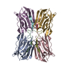 1ku8SC S: Starting model for refinement C: citing same article ( |
|---|---|
| Similar structure data |
-Links
 PDBj
PDBj- Assembly
| Deposited unit | 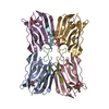
| ||||||||
|---|---|---|---|---|---|---|---|---|---|
| 1 |
| ||||||||
| Unit cell |
| ||||||||
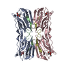
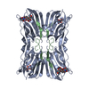
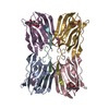
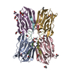
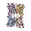
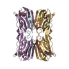
| Details | TETRAMER OF FOUR ALPHA CHAINS ASSOCIATED WITH FOUR BETA CHAINS. THIS ENTRY CONTAINS A COMPLETE TETRAMER |
-Components
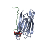
| #1: Protein | Mass: 14673.479 Da / Num. of mol.: 4 / Source method: isolated from a natural source / Source: (natural) Artocarpus integer (chempedak) / References: UniProt: P18670#2: Protein/peptide | Mass: 1872.064 Da / Num. of mol.: 4 / Source method: isolated from a natural source / Source: (natural) Artocarpus integer (chempedak) / References: UniProt: P18671#3: Sugar | ChemComp-MMA / 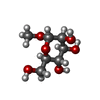  Type: D-saccharide / Mass: 194.182 Da / Num. of mol.: 4 / Source method: obtained synthetically / Formula: C7H14O6 Type: D-saccharide / Mass: 194.182 Da / Num. of mol.: 4 / Source method: obtained synthetically / Formula: C7H14O6#4: Water | ChemComp-HOH / |  Mass: 18.015 Da / Num. of mol.: 305 / Source method: isolated from a natural source / Formula: H2O Mass: 18.015 Da / Num. of mol.: 305 / Source method: isolated from a natural source / Formula: H2O |
|---|
-Experimental details
-Experiment
| Experiment | Method: X-RAY DIFFRACTION / Number of used crystals: 1 |
|---|
- Sample preparation
| Crystal | Density Matthews: 2.23 Å3/Da / Density % sol: 44.82 % | |||||||||||||||||||||||||||||||||||||||||||||||||
|---|---|---|---|---|---|---|---|---|---|---|---|---|---|---|---|---|---|---|---|---|---|---|---|---|---|---|---|---|---|---|---|---|---|---|---|---|---|---|---|---|---|---|---|---|---|---|---|---|---|---|
| Crystal grow | Temperature: 293 K / Method: vapor diffusion, hanging drop / pH: 7.5 Details: 20% PEG 8K, 10% ISOPROPANOL (V/V), 0.1 M HEPES, pH 7.5, VAPOR DIFFUSION, HANGING DROP, temperature 293K | |||||||||||||||||||||||||||||||||||||||||||||||||
| Crystal grow | *PLUS Temperature: 20 ℃ / pH: 7.4 / Method: vapor diffusion | |||||||||||||||||||||||||||||||||||||||||||||||||
| Components of the solutions | *PLUS
|
-Data collection
| Diffraction | Mean temperature: 100 K |
|---|---|
| Diffraction source | Source: SYNCHROTRON / Site: ESRF  / Beamline: BM14 / Wavelength: 0.984 Å / Beamline: BM14 / Wavelength: 0.984 Å |
| Detector | Type: MARRESEARCH / Detector: AREA DETECTOR / Date: Mar 2, 2000 |
| Radiation | Monochromator: MIRROR / Protocol: SINGLE WAVELENGTH / Monochromatic (M) / Laue (L): M / Scattering type: x-ray |
| Radiation wavelength | Wavelength: 0.984 Å / Relative weight: 1 |
| Reflection | Resolution: 2→33 Å / Num. obs: 38447 / % possible obs: 98.1 % / Observed criterion σ(I): -3 / Redundancy: 1.9 % / Biso Wilson estimate: 9.1 Å2 / Rsym value: 0.038 / Net I/σ(I): 9.5 |
| Reflection | *PLUS Highest resolution: 1.9 Å / Num. obs: 38468 / Num. measured all: 140888 / Rmerge(I) obs: 0.042 |
| Reflection shell | *PLUS % possible obs: 88.5 % / Rmerge(I) obs: 0.127 / Mean I/σ(I) obs: 3.7 |
- Processing
| Software |
| ||||||||||||||||||||||||||||||||||||||||||||||||||||||||||||
|---|---|---|---|---|---|---|---|---|---|---|---|---|---|---|---|---|---|---|---|---|---|---|---|---|---|---|---|---|---|---|---|---|---|---|---|---|---|---|---|---|---|---|---|---|---|---|---|---|---|---|---|---|---|---|---|---|---|---|---|---|---|
| Refinement | Method to determine structure: FOURIER SYNTHESIS Starting model: 1KU8 Resolution: 2→19.94 Å / Rfactor Rfree error: 0.008 / Data cutoff high absF: 1509605.77 / Data cutoff low absF: 0 / Isotropic thermal model: RESTRAINED / Cross valid method: THROUGHOUT / σ(F): 0 / Stereochemistry target values: Engh & Huber
| ||||||||||||||||||||||||||||||||||||||||||||||||||||||||||||
| Solvent computation | Solvent model: FLAT MODEL / Bsol: 41.8744 Å2 / ksol: 0.330889 e/Å3 | ||||||||||||||||||||||||||||||||||||||||||||||||||||||||||||
| Displacement parameters | Biso mean: 29.4 Å2
| ||||||||||||||||||||||||||||||||||||||||||||||||||||||||||||
| Refine analyze |
| ||||||||||||||||||||||||||||||||||||||||||||||||||||||||||||
| Refinement step | Cycle: LAST / Resolution: 2→19.94 Å
| ||||||||||||||||||||||||||||||||||||||||||||||||||||||||||||
| Refine LS restraints |
| ||||||||||||||||||||||||||||||||||||||||||||||||||||||||||||
| LS refinement shell | Resolution: 2→2.13 Å / Rfactor Rfree error: 0.028 / Total num. of bins used: 6
| ||||||||||||||||||||||||||||||||||||||||||||||||||||||||||||
| Xplor file |
| ||||||||||||||||||||||||||||||||||||||||||||||||||||||||||||
| Refinement | *PLUS Highest resolution: 2 Å / Lowest resolution: 20 Å / Rfactor Rfree: 0.235 / Rfactor Rwork: 0.191 | ||||||||||||||||||||||||||||||||||||||||||||||||||||||||||||
| Solvent computation | *PLUS | ||||||||||||||||||||||||||||||||||||||||||||||||||||||||||||
| Displacement parameters | *PLUS | ||||||||||||||||||||||||||||||||||||||||||||||||||||||||||||
| Refine LS restraints | *PLUS
| ||||||||||||||||||||||||||||||||||||||||||||||||||||||||||||
| LS refinement shell | *PLUS Rfactor Rfree: 0.324 / Rfactor Rwork: 0.275 |
