 Movie
Movie Controller


+ Open data

- Basic information
| Entry | Database: PDB / ID: 5it5 | ||||||
|---|---|---|---|---|---|---|---|
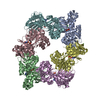
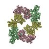
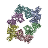
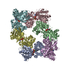
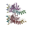
| Title | Thermus thermophilus PilB core ATPase region | ||||||
 Components Components | ATP binding motif-containing protein PilF | ||||||
 Keywords Keywords | TRANSPORT PROTEIN / ATPase / AAA+ / hexamer / Type IV pilus | ||||||
| Function / homology |  Function and homology information Function and homology informationATP hydrolysis activity / ATP binding / metal ion binding / plasma membrane / cytoplasm Similarity search - Function | ||||||
| Biological species |   Thermus thermophilus (bacteria) Thermus thermophilus (bacteria) | ||||||
| Method |  X-RAY DIFFRACTION / SYNCHROTRON / MOLECULAR REPLACEMENT / Resolution: 2.648 Å X-RAY DIFFRACTION / SYNCHROTRON / MOLECULAR REPLACEMENT / Resolution: 2.648 Å | ||||||
 Authors Authors | Mancl, J. / Robinson, H. / Black, W. / Yang, Z. / Schubot, F. | ||||||
| Funding support |  United States, 1items United States, 1items
| ||||||
 Citation Citation | Journal: Structure / Year: 2016 Title: Crystal Structure of a Type IV Pilus Assembly ATPase: Insights into the Molecular Mechanism of PilB from Thermus thermophilus. Authors: Mancl, J.M. / Black, W.P. / Robinson, H. / Yang, Z. / Schubot, F.D. | ||||||
| History |
|
- Structure visualization
| Structure viewer | Molecule: MolmilJmol/JSmol |
|---|
- Downloads & links
-Download
| PDBx/mmCIF format | 5it5.cif.gz | 453.9 KB | Display | PDBx/mmCIF format |
|---|---|---|---|---|
| PDB format | pdb5it5.ent.gz | 370.5 KB | Display | PDB format |
| PDBx/mmJSON format | 5it5.json.gz | Tree view | PDBx/mmJSON format | |
| Others |  Other downloads Other downloads |
-Validation report
| Arichive directory | https://data.pdbj.org/pub/pdb/validation_reports/it/5it5ftp://data.pdbj.org/pub/pdb/validation_reports/it/5it5 | HTTPS FTP |
|---|
-Related structure data
| Related structure data |  1p9rS S: Starting model for refinement |
|---|---|
| Similar structure data |

-Links
 PDBj
PDBj

- Assembly
| Deposited unit | 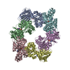
| ||||||||
|---|---|---|---|---|---|---|---|---|---|
| 1 |
| ||||||||
| Unit cell |
|
-Components
-Protein , 1 types, 6 molecules FABECD
| #1: Protein | Mass: 42526.992 Da / Num. of mol.: 6 / Fragment: unp residues 180-564 Source method: isolated from a genetically manipulated source Source: (gene. exp.) Thermus thermophilus (bacteria) / Gene: pilFProduction host: References: UniProt: Q8VRL1, UniProt: Q5SLC9*PLUS |
|---|
-Non-polymers , 5 types, 383 molecules 


| #2: Chemical | ChemComp-ZN /  Mass: 65.409 Da / Num. of mol.: 6 Mass: 65.409 Da / Num. of mol.: 6Source method: isolated from a genetically manipulated source Formula: Zn #3: Chemical | ChemComp-ATP / |  Mass: 507.181 Da / Num. of mol.: 1 Mass: 507.181 Da / Num. of mol.: 1Source method: isolated from a genetically manipulated source Formula: C10H16N5O13P3 / Comment: ATP, energy-carrying molecule*YM #4: Chemical | ChemComp-AGS /  Mass: 523.247 Da / Num. of mol.: 5 / Source method: isolated from a natural source / Formula: C10H16N5O12P3S / Comment: ATP-gamma-S, energy-carrying molecule analogue*YM Mass: 523.247 Da / Num. of mol.: 5 / Source method: isolated from a natural source / Formula: C10H16N5O12P3S / Comment: ATP-gamma-S, energy-carrying molecule analogue*YM#5: Chemical |  Mass: 24.305 Da / Num. of mol.: 2 / Source method: obtained synthetically / Formula: Mg Mass: 24.305 Da / Num. of mol.: 2 / Source method: obtained synthetically / Formula: Mg#6: Water | ChemComp-HOH / | Mass: 18.015 Da / Num. of mol.: 369 / Source method: isolated from a natural source / Formula: H2O |
|---|
-Experimental details
-Experiment
| Experiment | Method: X-RAY DIFFRACTION / Number of used crystals: 1 |
|---|
- Sample preparation
| Crystal | Density Matthews: 2.93 Å3/Da / Density % sol: 58.07 % |
|---|---|
| Crystal grow | Temperature: 293 K / Method: vapor diffusion, hanging drop / Details: Magnesium chloride, bis-tris, PEG 3350 |
-Data collection
| Diffraction | Mean temperature: 100 K |
|---|---|
| Diffraction source | Source: SYNCHROTRON / Site: NSLS / Beamline: X29A / Wavelength: 1.276396 Å |
| Detector | Type: ADSC QUANTUM 315 / Detector: CCD / Date: Jun 12, 2014 |
| Radiation | Protocol: SINGLE WAVELENGTH / Monochromatic (M) / Laue (L): M / Scattering type: x-ray |
| Radiation wavelength | Wavelength: 1.276396 Å / Relative weight: 1 |
| Reflection | Resolution: 2.648→65.29 Å / Num. obs: 87967 / % possible obs: 99.95 % / Redundancy: 2 % / Rmerge(I) obs: 0.1243 / Net I/σ(I): 5.83 |
| Reflection shell | Resolution: 2.65→2.74 Å |
- Processing
| Software |
| |||||||||||||||||||||||||||||||||||||||||||||||||||||||||||||||||||||||||||||||||||||||||||||||||||||||||||||||||||||||||||||||||||||||||||||||||||
|---|---|---|---|---|---|---|---|---|---|---|---|---|---|---|---|---|---|---|---|---|---|---|---|---|---|---|---|---|---|---|---|---|---|---|---|---|---|---|---|---|---|---|---|---|---|---|---|---|---|---|---|---|---|---|---|---|---|---|---|---|---|---|---|---|---|---|---|---|---|---|---|---|---|---|---|---|---|---|---|---|---|---|---|---|---|---|---|---|---|---|---|---|---|---|---|---|---|---|---|---|---|---|---|---|---|---|---|---|---|---|---|---|---|---|---|---|---|---|---|---|---|---|---|---|---|---|---|---|---|---|---|---|---|---|---|---|---|---|---|---|---|---|---|---|---|---|---|---|
| Refinement | Method to determine structure: MOLECULAR REPLACEMENT Starting model: 1P9R Resolution: 2.648→65.289 Å / SU ML: 0.37 / Cross valid method: THROUGHOUT / σ(F): 1.34 / Phase error: 31.4 / Stereochemistry target values: ML
| |||||||||||||||||||||||||||||||||||||||||||||||||||||||||||||||||||||||||||||||||||||||||||||||||||||||||||||||||||||||||||||||||||||||||||||||||||
| Solvent computation | Shrinkage radii: 0.9 Å / VDW probe radii: 1.11 Å / Solvent model: FLAT BULK SOLVENT MODEL | |||||||||||||||||||||||||||||||||||||||||||||||||||||||||||||||||||||||||||||||||||||||||||||||||||||||||||||||||||||||||||||||||||||||||||||||||||
| Displacement parameters | Biso max: 123.4 Å2 / Biso mean: 41.9005 Å2 / Biso min: 16.64 Å2 | |||||||||||||||||||||||||||||||||||||||||||||||||||||||||||||||||||||||||||||||||||||||||||||||||||||||||||||||||||||||||||||||||||||||||||||||||||
| Refinement step | Cycle: final / Resolution: 2.648→65.289 Å
| |||||||||||||||||||||||||||||||||||||||||||||||||||||||||||||||||||||||||||||||||||||||||||||||||||||||||||||||||||||||||||||||||||||||||||||||||||
| Refine LS restraints |
| |||||||||||||||||||||||||||||||||||||||||||||||||||||||||||||||||||||||||||||||||||||||||||||||||||||||||||||||||||||||||||||||||||||||||||||||||||
| LS refinement shell | Refine-ID: X-RAY DIFFRACTION / Total num. of bins used: 20
|
