 Movie
Movie Controller

[English] 日本語
 Yorodumi
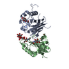
- PDB-5in2: Crystal structure of extra cellular Cu/Zn Superoxide Dismutase fr... -
+ Open data

- Basic information
| Entry | Database: PDB / ID: 5in2 | |||||||||
|---|---|---|---|---|---|---|---|---|---|---|
| Title | Crystal structure of extra cellular Cu/Zn Superoxide Dismutase from Onchocerca volvulus at 1.5 Angstrom; Insight into novel binding site and new inhibitors | |||||||||
 Components Components | Extracellular superoxide dismutase [Cu-Zn] | |||||||||
 Keywords Keywords | OXIDOREDUCTASE / Ec Ov-SOD / Onchcerca Volvulus / prodrug | |||||||||
| Function / homology |  Function and homology information Function and homology informationsuperoxide dismutase / superoxide dismutase activity / copper ion binding / extracellular region Similarity search - Function | |||||||||
| Biological species |  Onchocerca volvulus (invertebrata) Onchocerca volvulus (invertebrata) | |||||||||
| Method |  X-RAY DIFFRACTION / SYNCHROTRON / MOLECULAR REPLACEMENT / Resolution: 1.55 Å X-RAY DIFFRACTION / SYNCHROTRON / MOLECULAR REPLACEMENT / Resolution: 1.55 Å | |||||||||
 Authors Authors | Moustafa, A. / Betzel, C. / Perbandt, M. | |||||||||
| Funding support |  Egypt, Egypt,  Germany, 2items Germany, 2items
| |||||||||
 Citation Citation | Journal: To Be Published Title: Crystal structure of extracellular Cu/Zn Superoxide Dismutase from Onchocerca volvulus at 1.5 Angstrom; Insight into novel binding site and new inhibitors Authors: Moustafa, A. / Betzel, C. / Perbandt, M. | |||||||||
| History |
|
- Structure visualization
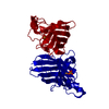
| Structure viewer | Molecule: MolmilJmol/JSmol |
|---|
- Downloads & links
-Download
| PDBx/mmCIF format | 5in2.cif.gz | 69.7 KB | Display | PDBx/mmCIF format |
|---|---|---|---|---|
| PDB format | pdb5in2.ent.gz | 50.6 KB | Display | PDB format |
| PDBx/mmJSON format | 5in2.json.gz | Tree view | PDBx/mmJSON format | |
| Others |  Other downloads Other downloads |
-Validation report
| Arichive directory | https://data.pdbj.org/pub/pdb/validation_reports/in/5in2ftp://data.pdbj.org/pub/pdb/validation_reports/in/5in2 | HTTPS FTP |
|---|
-Related structure data
| Related structure data |  3kbeS S: Starting model for refinement |
|---|---|
| Similar structure data |


-Links
 PDBj
PDBj
- Assembly
| Deposited unit | 
| ||||||||
|---|---|---|---|---|---|---|---|---|---|
| 1 | 
| ||||||||
| Unit cell |
|
-Components
-Protein , 1 types, 1 molecules A
| #1: Protein | Mass: 16097.852 Da / Num. of mol.: 1 Source method: isolated from a genetically manipulated source Source: (gene. exp.) Onchocerca volvulus (invertebrata) / Gene: sod-4, sod2 / Plasmid: pASKIBA16 / Production host:  |
|---|
-Non-polymers , 8 types, 99 molecules 


| #2: Chemical | ChemComp-GOL /  Mass: 92.094 Da / Num. of mol.: 1 / Source method: obtained synthetically / Formula: C3H8O3 Mass: 92.094 Da / Num. of mol.: 1 / Source method: obtained synthetically / Formula: C3H8O3 |
|---|---|
| #3: Chemical | ChemComp-ZN /  Mass: 65.409 Da / Num. of mol.: 1 / Source method: obtained synthetically / Formula: Zn Mass: 65.409 Da / Num. of mol.: 1 / Source method: obtained synthetically / Formula: Zn |
| #4: Chemical | ChemComp-CU /  Mass: 63.546 Da / Num. of mol.: 1 / Source method: obtained synthetically / Formula: Cu Mass: 63.546 Da / Num. of mol.: 1 / Source method: obtained synthetically / Formula: Cu |
| #5: Chemical | ChemComp-SO4 /  Mass: 96.063 Da / Num. of mol.: 1 / Source method: obtained synthetically / Formula: SO4 Mass: 96.063 Da / Num. of mol.: 1 / Source method: obtained synthetically / Formula: SO4 |
| #6: Chemical | ChemComp-CL /  Mass: 35.453 Da / Num. of mol.: 1 / Source method: obtained synthetically / Formula: Cl Mass: 35.453 Da / Num. of mol.: 1 / Source method: obtained synthetically / Formula: Cl |
| #7: Chemical | ChemComp-PGE /  Mass: 150.173 Da / Num. of mol.: 1 / Source method: obtained synthetically / Formula: C6H14O4 Mass: 150.173 Da / Num. of mol.: 1 / Source method: obtained synthetically / Formula: C6H14O4 |
| #8: Chemical | ChemComp-AZI /  Mass: 42.020 Da / Num. of mol.: 1 / Source method: obtained synthetically / Formula: N3 Mass: 42.020 Da / Num. of mol.: 1 / Source method: obtained synthetically / Formula: N3 |
| #9: Water | ChemComp-HOH / Mass: 18.015 Da / Num. of mol.: 92 / Source method: isolated from a natural source / Formula: H2O |
-Details
| Has protein modification | Y |
|---|
-Experimental details
-Experiment
| Experiment | Method: X-RAY DIFFRACTION / Number of used crystals: 1 |
|---|
- Sample preparation
| Crystal | Density Matthews: 2.4 Å3/Da / Density % sol: 48.8 % |
|---|---|
| Crystal grow | Temperature: 293 K / Method: vapor diffusion, sitting drop Details: 10 % w/v PEG 20 000, 20 % v/v PEG MME 550, 0.03 M of each ethylene glycol and 0.1 M MOPS/HEPES-Na pH 7.5 |
-Data collection
| Diffraction | Mean temperature: 100 K |
|---|---|
| Diffraction source | Source: SYNCHROTRON / Site: PETRA III, EMBL c/o DESY / Beamline: P13 (MX1) / Wavelength: 1.033 Å |
| Detector | Type: PSI PILATUS 6M / Detector: PIXEL / Date: Jun 23, 2015 |
| Radiation | Protocol: SINGLE WAVELENGTH / Monochromatic (M) / Laue (L): M / Scattering type: x-ray |
| Radiation wavelength | Wavelength: 1.033 Å / Relative weight: 1 |
| Reflection | Resolution: 1.55→25.292 Å / Num. obs: 22806 / % possible obs: 100 % / Redundancy: 8.7 % / Rmerge(I) obs: 0.075 / Net I/σ(I): 14.1 |
| Reflection shell | Highest resolution: 1.55 Å / Rmerge(I) obs: 0.42 |
- Processing
| Software |
| |||||||||||||||||||||||||||||||||||||||||||||||||||||||||||||||
|---|---|---|---|---|---|---|---|---|---|---|---|---|---|---|---|---|---|---|---|---|---|---|---|---|---|---|---|---|---|---|---|---|---|---|---|---|---|---|---|---|---|---|---|---|---|---|---|---|---|---|---|---|---|---|---|---|---|---|---|---|---|---|---|---|
| Refinement | Method to determine structure: MOLECULAR REPLACEMENT Starting model: 3KBE Resolution: 1.55→25.292 Å / SU ML: 0.14 / Cross valid method: FREE R-VALUE / σ(F): 1.53 / Phase error: 17.31
| |||||||||||||||||||||||||||||||||||||||||||||||||||||||||||||||
| Solvent computation | Shrinkage radii: 0.9 Å / VDW probe radii: 1.11 Å | |||||||||||||||||||||||||||||||||||||||||||||||||||||||||||||||
| Refinement step | Cycle: LAST / Resolution: 1.55→25.292 Å
| |||||||||||||||||||||||||||||||||||||||||||||||||||||||||||||||
| Refine LS restraints |
| |||||||||||||||||||||||||||||||||||||||||||||||||||||||||||||||
| LS refinement shell |
|
