 Movie
Movie Controller

[English] 日本語
 Yorodumi
- PDB-5il6: Crystal structure of the dehydratase domain of RzxB from Pseudomo... -
+ Open data

- Basic information
| Entry | Database: PDB / ID: 5il6 | ||||||
|---|---|---|---|---|---|---|---|
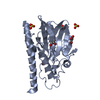
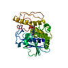
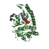
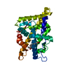
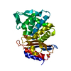
| Title | Crystal structure of the dehydratase domain of RzxB from Pseudomonas fluorescens | ||||||
 Components Components | Polyketide synthase/nonribosomal peptide synthetase hybrid RzxB | ||||||
 Keywords Keywords | HYDROLASE / polyketide / macrolactin / trans-AT / pKS / AT-less / dehydratase / rhizoxin | ||||||
| Function / homology |  Function and homology information Function and homology informationnonribosomal peptide biosynthetic process / polyketide biosynthetic process / DIM/DIP cell wall layer assembly / fatty acid synthase activity / ligase activity / phosphopantetheine binding / 3-oxoacyl-[acyl-carrier-protein] synthase activity / fatty acid biosynthetic process / oxidoreductase activity / plasma membrane / cytoplasm Similarity search - Function | ||||||
| Biological species |  Pseudomonas fluorescens (bacteria) Pseudomonas fluorescens (bacteria) | ||||||
| Method |  X-RAY DIFFRACTION / SYNCHROTRON / MOLECULAR REPLACEMENT / Resolution: 1.9 Å X-RAY DIFFRACTION / SYNCHROTRON / MOLECULAR REPLACEMENT / Resolution: 1.9 Å | ||||||
 Authors Authors | Jakob, R.P. / Herbst, D.A. / Maier, T. | ||||||
 Citation Citation | Journal: To Be Published Title: Crystal Structures of Dehydratase Domains from trans-AT Polyketide Biosynthetic Pathway Authors: Jakob, R.P. / Muller, R. / Herbst, D.A. / Maier, T. | ||||||
| History |
|
- Structure visualization
| Structure viewer | Molecule: MolmilJmol/JSmol |
|---|
- Downloads & links
-Download
| PDBx/mmCIF format | 5il6.cif.gz | 317.5 KB | Display | PDBx/mmCIF format |
|---|---|---|---|---|
| PDB format | pdb5il6.ent.gz | 265.6 KB | Display | PDB format |
| PDBx/mmJSON format | 5il6.json.gz | Tree view | PDBx/mmJSON format | |
| Others |  Other downloads Other downloads |
-Validation report
| Summary document | 5il6_validation.pdf.gz | 431.7 KB | Display | wwPDB validaton report |
|---|---|---|---|---|
| Full document | 5il6_full_validation.pdf.gz | 434.8 KB | Display | |
| Data in XML | 5il6_validation.xml.gz | 24.6 KB | Display | |
| Data in CIF | 5il6_validation.cif.gz | 35.8 KB | Display | |
| Arichive directory | https://data.pdbj.org/pub/pdb/validation_reports/il/5il6ftp://data.pdbj.org/pub/pdb/validation_reports/il/5il6 | HTTPS FTP |
-Related structure data
| Related structure data | 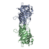 5hstC 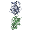 5hu7C 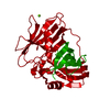 5j6oC  3kg9S S: Starting model for refinement C: citing same article ( |
|---|---|
| Similar structure data |


-Links
 PDBj
PDBj


- Assembly
| Deposited unit | 
| ||||||||
|---|---|---|---|---|---|---|---|---|---|
| 1 | 
| ||||||||
| 2 | 
| ||||||||
| Unit cell |
|
-Components
| #1: Protein | Mass: 33525.215 Da / Num. of mol.: 2 / Fragment: UNP residues 1231-1540 Source method: isolated from a genetically manipulated source Source: (gene. exp.) Pseudomonas fluorescens (strain Pf-5 / ATCC BAA-477) (bacteria)Strain: Pf-5 / ATCC BAA-477 / Gene: rzxB, PFL_2989 / Plasmid: pNIC28-Bsa4 / Production host: #2: Water | ChemComp-HOH / |  Mass: 18.015 Da / Num. of mol.: 384 / Source method: isolated from a natural source / Formula: H2O Mass: 18.015 Da / Num. of mol.: 384 / Source method: isolated from a natural source / Formula: H2O |
|---|
-Experimental details
-Experiment
| Experiment | Method: X-RAY DIFFRACTION / Number of used crystals: 1 |
|---|
- Sample preparation
| Crystal | Density Matthews: 2.65 Å3/Da / Density % sol: 53.51 % |
|---|---|
| Crystal grow | Temperature: 277 K / Method: vapor diffusion, hanging drop / Details: 0.1M BisTris 6.5, 0.2 M NaCl, 20% PEG6000 |
-Data collection
| Diffraction | Mean temperature: 100 K |
|---|---|
| Diffraction source | Source: SYNCHROTRON / Site: SLS  / Beamline: X06SA / Wavelength: 1 Å / Beamline: X06SA / Wavelength: 1 Å |
| Detector | Type: DECTRIS PILATUS 6M-F / Detector: PIXEL / Date: Aug 30, 2015 |
| Radiation | Protocol: SINGLE WAVELENGTH / Monochromatic (M) / Laue (L): M / Scattering type: x-ray |
| Radiation wavelength | Wavelength: 1 Å / Relative weight: 1 |
| Reflection | Resolution: 1.9→63.217 Å / Num. obs: 50913 / % possible obs: 98.1 % / Redundancy: 3.3 % / CC1/2: 0.997 / Rmerge(I) obs: 0.067 / Net I/σ(I): 13.1 |
| Reflection shell | Resolution: 1.9→2.01 Å / Redundancy: 3.2 % / Rmerge(I) obs: 0.482 / Mean I/σ(I) obs: 2.4 / % possible all: 97.3 |
- Processing
| Software |
| |||||||||||||||||||||||||||||||||||||||||||||||||||||||||||||||||||||||||||||||||||||||||||||||||||||||||||||||||||||||||||||||||||||||||||||||||||||||||||||||||||||||||||||||
|---|---|---|---|---|---|---|---|---|---|---|---|---|---|---|---|---|---|---|---|---|---|---|---|---|---|---|---|---|---|---|---|---|---|---|---|---|---|---|---|---|---|---|---|---|---|---|---|---|---|---|---|---|---|---|---|---|---|---|---|---|---|---|---|---|---|---|---|---|---|---|---|---|---|---|---|---|---|---|---|---|---|---|---|---|---|---|---|---|---|---|---|---|---|---|---|---|---|---|---|---|---|---|---|---|---|---|---|---|---|---|---|---|---|---|---|---|---|---|---|---|---|---|---|---|---|---|---|---|---|---|---|---|---|---|---|---|---|---|---|---|---|---|---|---|---|---|---|---|---|---|---|---|---|---|---|---|---|---|---|---|---|---|---|---|---|---|---|---|---|---|---|---|---|---|---|---|
| Refinement | Method to determine structure: MOLECULAR REPLACEMENT Starting model: 3KG9 Resolution: 1.9→63.217 Å / SU ML: 0.22 / Cross valid method: FREE R-VALUE / σ(F): 1.38 / Phase error: 21.9
| |||||||||||||||||||||||||||||||||||||||||||||||||||||||||||||||||||||||||||||||||||||||||||||||||||||||||||||||||||||||||||||||||||||||||||||||||||||||||||||||||||||||||||||||
| Solvent computation | Shrinkage radii: 0.9 Å / VDW probe radii: 1.11 Å | |||||||||||||||||||||||||||||||||||||||||||||||||||||||||||||||||||||||||||||||||||||||||||||||||||||||||||||||||||||||||||||||||||||||||||||||||||||||||||||||||||||||||||||||
| Refinement step | Cycle: LAST / Resolution: 1.9→63.217 Å
| |||||||||||||||||||||||||||||||||||||||||||||||||||||||||||||||||||||||||||||||||||||||||||||||||||||||||||||||||||||||||||||||||||||||||||||||||||||||||||||||||||||||||||||||
| Refine LS restraints |
| |||||||||||||||||||||||||||||||||||||||||||||||||||||||||||||||||||||||||||||||||||||||||||||||||||||||||||||||||||||||||||||||||||||||||||||||||||||||||||||||||||||||||||||||
| LS refinement shell |
| |||||||||||||||||||||||||||||||||||||||||||||||||||||||||||||||||||||||||||||||||||||||||||||||||||||||||||||||||||||||||||||||||||||||||||||||||||||||||||||||||||||||||||||||
| Refinement TLS params. | Method: refined / Refine-ID: X-RAY DIFFRACTION
| |||||||||||||||||||||||||||||||||||||||||||||||||||||||||||||||||||||||||||||||||||||||||||||||||||||||||||||||||||||||||||||||||||||||||||||||||||||||||||||||||||||||||||||||
| Refinement TLS group |
|
