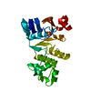
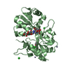
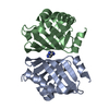
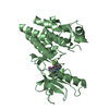
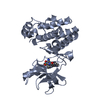
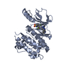
Entry Database : PDB / ID : 5ikbTitle Crystal structure of the kainate receptor GluK4 ligand binding domain in complex with kainate Glutamate receptor ionotropic, kainate 4,Glutamate receptor ionotropic, kainate 4 Keywords / / / Function / homology Function Domain/homology Component
/ / / / / / / / / / / / / / / / / / / / / / / / / / / / / / / / / / / / / / / / / / / / / / / / / / / / / / Biological species Rattus norvegicus (Norway rat)Method / / / Resolution : 2.05 Å Authors Kristensen, O. / Kristensen, L.B. / Frydenvang, K. / Kastrup, J.S. Funding support Organization Grant number Country The Lundbeck Foundation E.C. (FP7) Biostruct-X
Journal : Structure / Year : 2016Title : The Structure of a High-Affinity Kainate Receptor: GluK4 Ligand-Binding Domain Crystallized with Kainate.Authors : Kristensen, O. / Kristensen, L.B. / Mollerud, S. / Frydenvang, K. / Pickering, D.S. / Kastrup, J.S. History Deposition Mar 3, 2016 Deposition site / Processing site Revision 1.0 Aug 24, 2016 Provider / Type Revision 1.1 Sep 14, 2016 Group Revision 1.2 Aug 9, 2017 Group / Category / Item Revision 2.0 Mar 27, 2019 Group / Data collection / Source and taxonomy / Category / entity_src_genItem / _entity_src_gen.pdbx_host_org_cell_line / _entity_src_gen.pdbx_host_org_strainRevision 2.1 Jan 10, 2024 Group / Database references / Refinement descriptionCategory chem_comp_atom / chem_comp_bond ... chem_comp_atom / chem_comp_bond / database_2 / pdbx_initial_refinement_model Item / _database_2.pdbx_database_accession
Show all Show less
 Movie
Movie Controller

 Yorodumi
 Open data
 Basic information
 Components
 Keywords
 Function and homology information
 X-RAY DIFFRACTION /
  Authors
 Denmark, 2items
  Citation
 Structure visualization
 Downloads & links
 Other downloads

 PDBj
PDBj

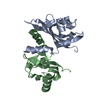
 Assembly
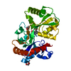
 Trichoplusia ni (cabbage looper) / References: UniProt: Q01812
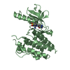
 Mass: 213.230 Da / Num. of mol.: 1 / Source method: obtained synthetically / Formula: C10H15NO4 / Comment: neurotransmitter, agonist*YM
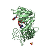
 Mass: 92.094 Da / Num. of mol.: 2 / Source method: obtained synthetically / Formula: C3H8O3
 Mass: 18.015 Da / Num. of mol.: 220 / Source method: isolated from a natural source / Formula: H2O
 Sample preparation
 / Beamline: MASSIF-1 / Wavelength: 0.966 Å
 Processing