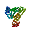
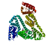
Journal: J.Med.Chem. / Year: 2016 Title: X-ray Structure Analysis of Indazolium trans-[Tetrachlorobis(1H-indazole)ruthenate(III)] (KP1019) Bound to Human Serum Albumin Reveals Two Ruthenium Binding Sites and Provides Insights into ...Title: X-ray Structure Analysis of Indazolium trans-[Tetrachlorobis(1H-indazole)ruthenate(III)] (KP1019) Bound to Human Serum Albumin Reveals Two Ruthenium Binding Sites and Provides Insights into the Drug Binding Mechanism. Authors: Bijelic, A. / Theiner, S. / Keppler, B.K. / Rompel, A.
Resolution: 3.2→45.844 Å / SU ML: 0.46 / Cross valid method: FREE R-VALUE / σ(F): 1.93 / Phase error: 30.21 Details: During processing the data was cut at 2.93 A due to suitable values for I/sigma, CC1/2 etc. However, during refinement a great drop in data completeness beginning at >3.2 A was recognized ...Details: During processing the data was cut at 2.93 A due to suitable values for I/sigma, CC1/2 etc. However, during refinement a great drop in data completeness beginning at >3.2 A was recognized and in addition the FOM was too small and the expected R-values were ridicilously high. The result was a blurry and strange looking map. Therefore, a 3.2 A cutoff was set during refinement.
Rfactor
Num. reflection
% reflection
Rfree
0.2615
959
4.95 %
Rwork
0.2441
-
-
obs
0.245
10501
95.97 %
Solvent computation
Shrinkage radii: 0.9 Å / VDW probe radii: 1.11 Å
Refinement step
Cycle: LAST / Resolution: 3.2→45.844 Å
Protein
Nucleic acid
Ligand
Solvent
Total
Num. atoms
4583
0
90
9
4682
Refine LS restraints
Refine-ID
Type
Dev ideal
Number
X-RAY DIFFRACTION
f_bond_d
0.016
4783
X-RAY DIFFRACTION
f_angle_d
0.413
6427
X-RAY DIFFRACTION
f_dihedral_angle_d
10.637
3007
X-RAY DIFFRACTION
f_chiral_restr
0.034
707
X-RAY DIFFRACTION
f_plane_restr
0.003
827
LS refinement shell
Resolution (Å)
Rfactor Rfree
Num. reflection Rfree
Rfactor Rwork
Num. reflection Rwork
Refine-ID
% reflection obs (%)
3.2-3.3687
0.3783
133
0.3618
2619
X-RAY DIFFRACTION
96
3.3687-3.5797
0.3423
130
0.3448
2549
X-RAY DIFFRACTION
93
3.5797-3.8559
0.3563
136
0.2967
2632
X-RAY DIFFRACTION
96
3.8559-4.2437
0.255
143
0.2566
2667
X-RAY DIFFRACTION
97
4.2437-4.8572
0.2167
137
0.2197
2692
X-RAY DIFFRACTION
98
4.8572-6.1172
0.2921
136
0.2362
2590
X-RAY DIFFRACTION
96
6.1172-45.8483
0.1919
144
0.1764
2648
X-RAY DIFFRACTION
97
+
About Yorodumi
-
News
-
Feb 9, 2022. New format data for meta-information of EMDB entries
New format data for meta-information of EMDB entries
Version 3 of the EMDB header file is now the official format.
The previous official version 1.9 will be removed from the archive.
In the structure databanks used in Yorodumi, some data are registered as the other names, "COVID-19 virus" and "2019-nCoV". Here are the details of the virus and the list of structure data.
Jan 31, 2019. EMDB accession codes are about to change! (news from PDBe EMDB page)
EMDB accession codes are about to change! (news from PDBe EMDB page)
The allocation of 4 digits for EMDB accession codes will soon come to an end. Whilst these codes will remain in use, new EMDB accession codes will include an additional digit and will expand incrementally as the available range of codes is exhausted. The current 4-digit format prefixed with “EMD-” (i.e. EMD-XXXX) will advance to a 5-digit format (i.e. EMD-XXXXX), and so on. It is currently estimated that the 4-digit codes will be depleted around Spring 2019, at which point the 5-digit format will come into force.
The EM Navigator/Yorodumi systems omit the EMD- prefix.
Related info.:Q: What is EMD? / ID/Accession-code notation in Yorodumi/EM Navigator
Yorodumi is a browser for structure data from EMDB, PDB, SASBDB, etc.
This page is also the successor to EM Navigator detail page, and also detail information page/front-end page for Omokage search.
The word "yorodu" (or yorozu) is an old Japanese word meaning "ten thousand". "mi" (miru) is to see.
Related info.:EMDB / PDB / SASBDB / Comparison of 3 databanks / Yorodumi Search / Aug 31, 2016. New EM Navigator & Yorodumi / Yorodumi Papers / Jmol/JSmol / Function and homology information / Changes in new EM Navigator and Yorodumi
 Movie
Movie Controller


 Open data
 Basic information
 Components
 Keywords
 Function and homology information
 Homo sapiens (human)
 X-RAY DIFFRACTION /
  Authors
 Austria, 1items
  Citation
 Structure visualization
 Downloads & links
 Other downloads
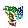


 PDBj
PDBj


 Assembly
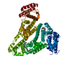

 Mass: 101.070 Da / Num. of mol.: 2 / Source method: obtained synthetically / Formula: Ru
 Mass: 228.371 Da / Num. of mol.: 6 / Source method: obtained synthetically / Formula: C14H28O2
 Mass: 18.015 Da / Num. of mol.: 9 / Source method: isolated from a natural source / Formula: H2O
 Sample preparation
 / Beamline: ID23-1 / Wavelength: 0.973 Å
 Processing