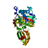
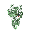
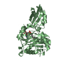
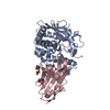
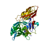
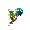
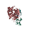
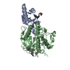
- PDB-5iby: Crystal structure of Enterococcus faecalis lipoate-protein ligase... -
+
Open data
ID or keywords:
Loading...
-
Basic information
Entry
Database: PDB / ID: 5iby
Title
Crystal structure of Enterococcus faecalis lipoate-protein ligase A (lplA-2) in complex with lipoic acid
Components
Lipoate--protein ligase
Keywords
LIGASE / TRANSFERASE / PROTEIN-SUBSTRATE COMPLEX
Function / homology
Function and homology information
lipoyltransferase activity / lipoate-protein ligase / lipoate-protein ligase activity / protein lipoylation / ATP binding / cytoplasm Similarity search - Function
Lipoate protein ligase, C-terminal / Bacterial lipoate protein ligase C-terminus / Lipoyltransferase/lipoate-protein ligase / Lipoyl protein ligase A/B catalytic domain / CO dehydrogenase flavoprotein, C-terminal domain / Biotinyl protein ligase (BPL) and lipoyl protein ligase (LPL) catalytic domain profile. / Biotin/lipoate A/B protein ligase family / Biotinyl protein ligase (BPL) and lipoyl protein ligase (LPL), catalytic domain / Bira Bifunctional Protein; Domain 2 / BirA Bifunctional Protein; domain 2 ...Lipoate protein ligase, C-terminal / Bacterial lipoate protein ligase C-terminus / Lipoyltransferase/lipoate-protein ligase / Lipoyl protein ligase A/B catalytic domain / CO dehydrogenase flavoprotein, C-terminal domain / Biotinyl protein ligase (BPL) and lipoyl protein ligase (LPL) catalytic domain profile. / Biotin/lipoate A/B protein ligase family / Biotinyl protein ligase (BPL) and lipoyl protein ligase (LPL), catalytic domain / Bira Bifunctional Protein; Domain 2 / BirA Bifunctional Protein; domain 2 / Enolase-like; domain 1 / Class II Aminoacyl-tRNA synthetase/Biotinyl protein ligase (BPL) and lipoyl protein ligase (LPL) / 2-Layer Sandwich / Alpha Beta Similarity search - Domain/homology
Mass: 18.015 Da / Num. of mol.: 279 / Source method: isolated from a natural source / Formula: H2O
-
Experimental details
-
Experiment
Experiment
Method: X-RAY DIFFRACTION / Number of used crystals: 1
-
Sample preparation
Crystal
Density Matthews: 2.1 Å3/Da / Density % sol: 41.3 % / Mosaicity: 0.444 °
Crystal grow
Temperature: 289 K / Method: vapor diffusion, hanging drop / pH: 5.25 Details: 1.5 UL PROTEIN + 1.5 UL BUFFER (25% PEG3350, 0.1 M SODIUM CACODYLATE, 0.2 M SODIUM CHLORIDE, PH 5.25)
Resolution: 1.85→21.5 Å / Cor.coef. Fo:Fc: 0.965 / Cor.coef. Fo:Fc free: 0.939 / WRfactor Rfree: 0.2139 / WRfactor Rwork: 0.1663 / FOM work R set: 0.8639 / SU B: 5.881 / SU ML: 0.093 / SU R Cruickshank DPI: 0.1369 / SU Rfree: 0.1314 / Cross valid method: THROUGHOUT / σ(F): 0 / ESU R: 0.137 / ESU R Free: 0.131 / Stereochemistry target values: MAXIMUM LIKELIHOOD Details: HYDROGENS HAVE BEEN ADDED IN THE RIDING POSITIONS U VALUES : WITH TLS ADDED
Rfactor
Num. reflection
% reflection
Selection details
Rfree
0.2103
1443
5.1 %
RANDOM
Rwork
0.1641
-
-
-
obs
0.1664
27075
99.4 %
-
Solvent computation
Ion probe radii: 0.7 Å / Shrinkage radii: 0.7 Å / VDW probe radii: 1.2 Å / Solvent model: MASK
In the structure databanks used in Yorodumi, some data are registered as the other names, "COVID-19 virus" and "2019-nCoV". Here are the details of the virus and the list of structure data.
Jan 31, 2019. EMDB accession codes are about to change! (news from PDBe EMDB page)
EMDB accession codes are about to change! (news from PDBe EMDB page)
The allocation of 4 digits for EMDB accession codes will soon come to an end. Whilst these codes will remain in use, new EMDB accession codes will include an additional digit and will expand incrementally as the available range of codes is exhausted. The current 4-digit format prefixed with “EMD-” (i.e. EMD-XXXX) will advance to a 5-digit format (i.e. EMD-XXXXX), and so on. It is currently estimated that the 4-digit codes will be depleted around Spring 2019, at which point the 5-digit format will come into force.
The EM Navigator/Yorodumi systems omit the EMD- prefix.
Related info.:Q: What is EMD? / ID/Accession-code notation in Yorodumi/EM Navigator
Yorodumi is a browser for structure data from EMDB, PDB, SASBDB, etc.
This page is also the successor to EM Navigator detail page, and also detail information page/front-end page for Omokage search.
The word "yorodu" (or yorozu) is an old Japanese word meaning "ten thousand". "mi" (miru) is to see.
Related info.:EMDB / PDB / SASBDB / Comparison of 3 databanks / Yorodumi Search / Aug 31, 2016. New EM Navigator & Yorodumi / Yorodumi Papers / Jmol/JSmol / Function and homology information / Changes in new EM Navigator and Yorodumi
 Movie
Movie Controller

 Yorodumi
 Open data
 Basic information
 Components
 Keywords
 Function and homology information
 Enterococcus faecalis (bacteria)
 X-RAY DIFFRACTION /
  Authors
 Citation
 Structure visualization
 Downloads & links
 Other downloads
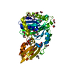


 PDBj
PDBj
 Assembly
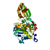
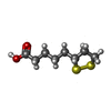
 Mass: 206.326 Da / Num. of mol.: 1 / Source method: obtained synthetically / Formula: C8H14O2S2
 Mass: 18.015 Da / Num. of mol.: 279 / Source method: isolated from a natural source / Formula: H2O
 Sample preparation
 / Beamline: 19-BM / Wavelength: 0.979054 Å
 Processing