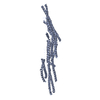
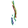
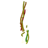
Entry Database : PDB / ID : 5i6rTitle Crystal Structure of srGAP2 F-BARx WT Form-1 SLIT-ROBO Rho GTPase-activating protein 2 Keywords / / / / Function / homology Function Domain/homology Component
/ / / / / / / / / / / / / / / / / / / / / / / / / / / / / / / / / / / / / / / / / / / / / / / / / / / / / / / / / / / / / / / / / / / / / / / / Biological species Homo sapiens (human)Method / / / Resolution : 2.15 Å Authors Sporny, M. / Guez-Haddad, J. / Isupov, M.N. / Opatowsky, Y. Funding support Organization Grant number Country ISF 182/10 and 1425/15 BSF 2013310
Journal : To Be Published Title : Structural Basis for srGAP2 Membrane Interactions, and Antagonism by the Human Specific Paralog srGAP2CAuthors : Sporny, M. / Guez-Haddad, J. / Isupov, M.N. / Opatowsky, Y. History Deposition Feb 16, 2016 Deposition site / Processing site Revision 1.0 Aug 30, 2017 Provider / Type Revision 1.1 Oct 16, 2019 Group / Category Revision 1.2 May 8, 2024 Group / Database references / Category / chem_comp_bond / database_2Item / _database_2.pdbx_database_accession
Show all Show less
 Movie
Movie Controller


 Open data
 Basic information
 Components
 Keywords
 Function and homology information
 Homo sapiens (human)
 X-RAY DIFFRACTION /
  Authors
 Israel, 2items
  Citation
 Structure visualization
 Downloads & links
 Other downloads


 PDBj
PDBj


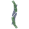
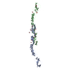
 Assembly
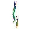


 Mass: 134.087 Da / Num. of mol.: 1 / Source method: obtained synthetically / Formula: C4H6O5
 Mass: 59.044 Da / Num. of mol.: 2 / Source method: obtained synthetically / Formula: C2H3O2
 Mass: 150.173 Da / Num. of mol.: 1 / Source method: obtained synthetically / Formula: C6H14O4
 Mass: 18.015 Da / Num. of mol.: 252 / Source method: isolated from a natural source / Formula: H2O
 Sample preparation
 / Beamline: 14.1 / Wavelength: 0.918 Å
 Processing