 Movie
Movie Controller


+ Open data

- Basic information
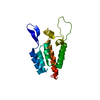
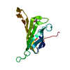
| Entry | Database: PDB / ID: 5i2x | ||||||
|---|---|---|---|---|---|---|---|
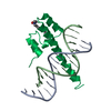
| Title | Crystal Structure of TPP1 K170del | ||||||
 Components Components | Adrenocortical dysplasia protein homolog | ||||||
 Keywords Keywords | PROTEIN BINDING / OB fold | ||||||
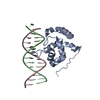
| Function / homology |  Function and homology information Function and homology informationtelomere assembly / segmentation / urogenital system development / protection from non-homologous end joining at telomere / establishment of protein localization to telomere / regulation of establishment of protein localization to telomere / telomerase inhibitor activity / shelterin complex / Telomere C-strand synthesis initiation / Telomere C-strand (Lagging Strand) Synthesis ...telomere assembly / segmentation / urogenital system development / protection from non-homologous end joining at telomere / establishment of protein localization to telomere / regulation of establishment of protein localization to telomere / telomerase inhibitor activity / shelterin complex / Telomere C-strand synthesis initiation / Telomere C-strand (Lagging Strand) Synthesis / embryonic limb morphogenesis / nuclear telomere cap complex / telomere capping / Polymerase switching on the C-strand of the telomere / telomerase holoenzyme complex / Processive synthesis on the C-strand of the telomere / Removal of the Flap Intermediate from the C-strand / protein localization to chromosome, telomeric region / telomeric repeat DNA binding / negative regulation of telomere maintenance via telomerase / positive regulation of telomere maintenance / Telomere Extension By Telomerase / telomere maintenance via telomerase / DNA polymerase binding / Packaging Of Telomere Ends / Recognition and association of DNA glycosylase with site containing an affected purine / Cleavage of the damaged purine / Recognition and association of DNA glycosylase with site containing an affected pyrimidine / Cleavage of the damaged pyrimidine / telomere maintenance / Inhibition of DNA recombination at telomere / Meiotic synapsis / skeletal system development / intracellular protein transport / DNA Damage/Telomere Stress Induced Senescence / chromosome, telomeric region / nuclear body / protein-containing complex binding / nucleoplasm Similarity search - Function | ||||||
| Biological species |  Homo sapiens (human) Homo sapiens (human) | ||||||
| Method |  X-RAY DIFFRACTION / SYNCHROTRON / MOLECULAR REPLACEMENT / Resolution: 3 Å X-RAY DIFFRACTION / SYNCHROTRON / MOLECULAR REPLACEMENT / Resolution: 3 Å | ||||||
 Authors Authors | Nandakumar, J. / Smith, E. | ||||||
| Funding support |  United States, 1items United States, 1items
| ||||||
 Citation Citation | Journal: Proc.Natl.Acad.Sci.USA / Year: 2016 Title: Structural and functional consequences of a disease mutation in the telomere protein TPP1. Authors: Bisht, K. / Smith, E.M. / Tesmer, V.M. / Nandakumar, J. | ||||||
| History |
|
- Structure visualization
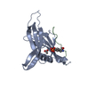
| Structure viewer | Molecule: MolmilJmol/JSmol |
|---|
- Downloads & links
-Download
| PDBx/mmCIF format | 5i2x.cif.gz | 69.7 KB | Display | PDBx/mmCIF format |
|---|---|---|---|---|
| PDB format | pdb5i2x.ent.gz | 50.7 KB | Display | PDB format |
| PDBx/mmJSON format | 5i2x.json.gz | Tree view | PDBx/mmJSON format | |
| Others |  Other downloads Other downloads |
-Validation report
| Arichive directory | https://data.pdbj.org/pub/pdb/validation_reports/i2/5i2xftp://data.pdbj.org/pub/pdb/validation_reports/i2/5i2x | HTTPS FTP |
|---|
-Related structure data
| Related structure data |  5i2yC  2i46S S: Starting model for refinement C: citing same article ( |
|---|---|
| Similar structure data |


-Links
 PDBj
PDBj


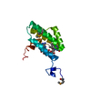
- Assembly
| Deposited unit | 
| |||||||||||||||||||||||||||||||||||||||||||||||||||||||||||||||||||||||||||||||||||||||||||||||||||||||||||||||||||||||||||||||||||||||||||||||||||||||||||||||||||||||||||||||||||||||||||||||||
|---|---|---|---|---|---|---|---|---|---|---|---|---|---|---|---|---|---|---|---|---|---|---|---|---|---|---|---|---|---|---|---|---|---|---|---|---|---|---|---|---|---|---|---|---|---|---|---|---|---|---|---|---|---|---|---|---|---|---|---|---|---|---|---|---|---|---|---|---|---|---|---|---|---|---|---|---|---|---|---|---|---|---|---|---|---|---|---|---|---|---|---|---|---|---|---|---|---|---|---|---|---|---|---|---|---|---|---|---|---|---|---|---|---|---|---|---|---|---|---|---|---|---|---|---|---|---|---|---|---|---|---|---|---|---|---|---|---|---|---|---|---|---|---|---|---|---|---|---|---|---|---|---|---|---|---|---|---|---|---|---|---|---|---|---|---|---|---|---|---|---|---|---|---|---|---|---|---|---|---|---|---|---|---|---|---|---|---|---|---|---|---|---|---|---|
| 1 | 
| |||||||||||||||||||||||||||||||||||||||||||||||||||||||||||||||||||||||||||||||||||||||||||||||||||||||||||||||||||||||||||||||||||||||||||||||||||||||||||||||||||||||||||||||||||||||||||||||||
| 2 | 
| |||||||||||||||||||||||||||||||||||||||||||||||||||||||||||||||||||||||||||||||||||||||||||||||||||||||||||||||||||||||||||||||||||||||||||||||||||||||||||||||||||||||||||||||||||||||||||||||||
| Unit cell |
| |||||||||||||||||||||||||||||||||||||||||||||||||||||||||||||||||||||||||||||||||||||||||||||||||||||||||||||||||||||||||||||||||||||||||||||||||||||||||||||||||||||||||||||||||||||||||||||||||
| Components on special symmetry positions |
| |||||||||||||||||||||||||||||||||||||||||||||||||||||||||||||||||||||||||||||||||||||||||||||||||||||||||||||||||||||||||||||||||||||||||||||||||||||||||||||||||||||||||||||||||||||||||||||||||
| Noncrystallographic symmetry (NCS) | NCS domain:
NCS domain segments: Component-ID: 1 / Refine code: 4
NCS ensembles :
NCS oper:
|
-Components
| #1: Protein | Mass: 17429.354 Da / Num. of mol.: 2 / Fragment: OB domain / Mutation: deletion of amino acid K170 Source method: isolated from a genetically manipulated source Source: (gene. exp.) Homo sapiens (human) / Gene: ACD, PIP1, PTOP, TINT1, TPP1 / Plasmid: pET-Smt3 / Production host:  #2: Water | ChemComp-HOH / |  Mass: 18.015 Da / Num. of mol.: 55 / Source method: isolated from a natural source / Formula: H2O Mass: 18.015 Da / Num. of mol.: 55 / Source method: isolated from a natural source / Formula: H2O |
|---|
-Experimental details
-Experiment
| Experiment | Method: X-RAY DIFFRACTION / Number of used crystals: 1 |
|---|
- Sample preparation
| Crystal | Density Matthews: 4.34 Å3/Da / Density % sol: 71.65 % / Mosaicity: 0.276 ° |
|---|---|
| Crystal grow | Temperature: 289 K / Method: vapor diffusion, hanging drop / pH: 8 / Details: 3.6 M sodium formate, 100 mM Tris-Cl pH 8.0 |
-Data collection
| Diffraction | Mean temperature: 100 K | |||||||||||||||||||||||||||||||||||||||||||||||||||||||
|---|---|---|---|---|---|---|---|---|---|---|---|---|---|---|---|---|---|---|---|---|---|---|---|---|---|---|---|---|---|---|---|---|---|---|---|---|---|---|---|---|---|---|---|---|---|---|---|---|---|---|---|---|---|---|---|---|
| Diffraction source | Source: SYNCHROTRON / Site: APS / Beamline: 21-ID-D / Wavelength: 1.03829 Å | |||||||||||||||||||||||||||||||||||||||||||||||||||||||
| Detector | Type: MAR scanner 300 mm plate / Detector: IMAGE PLATE / Date: Jun 16, 2014 | |||||||||||||||||||||||||||||||||||||||||||||||||||||||
| Radiation | Monochromator: Si(111) / Protocol: SINGLE WAVELENGTH / Monochromatic (M) / Laue (L): M / Scattering type: x-ray | |||||||||||||||||||||||||||||||||||||||||||||||||||||||
| Radiation wavelength | Wavelength: 1.03829 Å / Relative weight: 1 | |||||||||||||||||||||||||||||||||||||||||||||||||||||||
| Reflection | Resolution: 3→50 Å / Num. obs: 12617 / % possible obs: 99.7 % / Redundancy: 6.9 % / Rmerge(I) obs: 0.148 / Rpim(I) all: 0.059 / Rrim(I) all: 0.16 / Χ2: 1.496 / Net I/av σ(I): 14.929 / Net I/σ(I): 7.1 / Num. measured all: 86466 | |||||||||||||||||||||||||||||||||||||||||||||||||||||||
| Reflection shell |
|
- Processing
| Software |
| ||||||||||||||||||||||||||||||||||||||||||||||||||||||||||||
|---|---|---|---|---|---|---|---|---|---|---|---|---|---|---|---|---|---|---|---|---|---|---|---|---|---|---|---|---|---|---|---|---|---|---|---|---|---|---|---|---|---|---|---|---|---|---|---|---|---|---|---|---|---|---|---|---|---|---|---|---|---|
| Refinement | Method to determine structure: MOLECULAR REPLACEMENT Starting model: 2I46 Resolution: 3→50 Å / Cor.coef. Fo:Fc: 0.925 / Cor.coef. Fo:Fc free: 0.897 / SU B: 12.346 / SU ML: 0.222 / Cross valid method: THROUGHOUT / σ(F): 0 / ESU R: 0.578 / ESU R Free: 0.312 / Details: U VALUES : REFINED INDIVIDUALLY
| ||||||||||||||||||||||||||||||||||||||||||||||||||||||||||||
| Solvent computation | Ion probe radii: 0.8 Å / Shrinkage radii: 0.8 Å / VDW probe radii: 1.2 Å | ||||||||||||||||||||||||||||||||||||||||||||||||||||||||||||
| Displacement parameters | Biso max: 129.16 Å2 / Biso mean: 37.111 Å2 / Biso min: 2 Å2
| ||||||||||||||||||||||||||||||||||||||||||||||||||||||||||||
| Refinement step | Cycle: final / Resolution: 3→50 Å
| ||||||||||||||||||||||||||||||||||||||||||||||||||||||||||||
| Refine LS restraints |
| ||||||||||||||||||||||||||||||||||||||||||||||||||||||||||||
| Refine LS restraints NCS | Dom-ID: 1 / Auth asym-ID: A / Refine-ID: X-RAY DIFFRACTION
| ||||||||||||||||||||||||||||||||||||||||||||||||||||||||||||
| LS refinement shell | Resolution: 3→3.078 Å / Total num. of bins used: 20
|
