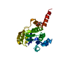
[English] 日本語
Yorodumi
- PDB-1yrk: The C2 Domain of PKC is a new Phospho-Tyrosine Binding Domain -

+
Open data


ID or keywords:

Loading...

-
Basic information

Entry
Database: PDB / ID: 1yrk
TitleThe C2 Domain of PKC is a new Phospho-Tyrosine Binding Domain
Components
  • 13-residue peptide
  • Protein kinase C, delta type
KeywordsPROTEIN BINDING / C2 domain
Function / homology
Function and homology information


positive regulation of phospholipid scramblase activity / positive regulation of glucosylceramide catabolic process / positive regulation of sphingomyelin catabolic process / diacylglycerol-dependent, calcium-independent serine/threonine kinase activity / regulation of ceramide biosynthetic process / HuR (ELAVL1) binds and stabilizes mRNA / endolysosome / positive regulation of ceramide biosynthetic process / negative regulation of peptidyl-tyrosine phosphorylation / termination of signal transduction ...positive regulation of phospholipid scramblase activity / positive regulation of glucosylceramide catabolic process / positive regulation of sphingomyelin catabolic process / diacylglycerol-dependent, calcium-independent serine/threonine kinase activity / regulation of ceramide biosynthetic process / HuR (ELAVL1) binds and stabilizes mRNA / endolysosome / positive regulation of ceramide biosynthetic process / negative regulation of peptidyl-tyrosine phosphorylation / termination of signal transduction / negative regulation of filopodium assembly / DAG and IP3 signaling / protein kinase C / cellular response to hydroperoxide / negative regulation of glial cell apoptotic process / diacylglycerol-dependent serine/threonine kinase activity / Effects of PIP2 hydrolysis / negative regulation of actin filament polymerization / negative regulation of platelet aggregation / positive regulation of endodeoxyribonuclease activity / neutrophil activation / regulation of signaling receptor activity / cellular response to angiotensin / Calmodulin induced events / intrinsic apoptotic signaling pathway in response to oxidative stress / Fc-gamma receptor signaling pathway involved in phagocytosis / activation of protein kinase activity / B cell proliferation / Apoptotic cleavage of cellular proteins / insulin receptor substrate binding / RHO GTPases Activate NADPH Oxidases / immunoglobulin mediated immune response / Role of phospholipids in phagocytosis / positive regulation of protein dephosphorylation / enzyme activator activity / regulation of mRNA stability / negative regulation of insulin receptor signaling pathway / SHC1 events in ERBB2 signaling / post-translational protein modification / cell chemotaxis / positive regulation of superoxide anion generation / VEGFR2 mediated cell proliferation / negative regulation of MAP kinase activity / negative regulation of protein binding / regulation of actin cytoskeleton organization / positive regulation of apoptotic signaling pathway / peptidyl-threonine phosphorylation / non-specific protein-tyrosine kinase / non-membrane spanning protein tyrosine kinase activity / CLEC7A (Dectin-1) signaling / nuclear matrix / negative regulation of inflammatory response / G alpha (z) signalling events / cellular response to hydrogen peroxide / positive regulation of protein import into nucleus / cellular response to UV / KEAP1-NFE2L2 pathway / Interferon gamma signaling / azurophil granule lumen / cellular senescence / cell-cell junction / peptidyl-serine phosphorylation / protein stabilization / protein kinase activity / intracellular signal transduction / defense response to bacterium / cell cycle / protein phosphorylation / protein serine kinase activity / protein serine/threonine kinase activity / apoptotic process / DNA damage response / Neutrophil degranulation / protein kinase binding / perinuclear region of cytoplasm / enzyme binding / endoplasmic reticulum / signal transduction / mitochondrion / extracellular exosome / extracellular region / nucleoplasm / ATP binding / nucleus / metal ion binding / plasma membrane / cytoplasm / cytosol
Similarity search - Function
Novel protein kinase C delta, catalytic domain / Protein kinase C, delta / Protein kinase C delta/epsilon/eta/theta, C2 domain / Protein kinase C, delta/epsilon/eta/theta types / Protein kinase, C-terminal / Protein kinase C terminal domain / Diacylglycerol/phorbol-ester binding / C2 domain / Phorbol esters/diacylglycerol binding domain (C1 domain) / Zinc finger phorbol-ester/DAG-type signature. ...Novel protein kinase C delta, catalytic domain / Protein kinase C, delta / Protein kinase C delta/epsilon/eta/theta, C2 domain / Protein kinase C, delta/epsilon/eta/theta types / Protein kinase, C-terminal / Protein kinase C terminal domain / Diacylglycerol/phorbol-ester binding / C2 domain / Phorbol esters/diacylglycerol binding domain (C1 domain) / Zinc finger phorbol-ester/DAG-type signature. / Zinc finger phorbol-ester/DAG-type profile. / Protein kinase C conserved region 1 (C1) domains (Cysteine-rich domains) / Protein kinase C-like, phorbol ester/diacylglycerol-binding domain / C2 domain / C2 domain profile. / C1-like domain superfamily / Extension to Ser/Thr-type protein kinases / AGC-kinase, C-terminal / AGC-kinase C-terminal domain profile. / C2 domain superfamily / Serine/threonine-protein kinase, active site / Serine/Threonine protein kinases active-site signature. / Protein kinase domain / Serine/Threonine protein kinases, catalytic domain / Protein kinase, ATP binding site / Protein kinases ATP-binding region signature. / Protein kinase domain profile. / Protein kinase domain / Protein kinase-like domain superfamily / Immunoglobulin-like / Sandwich / Mainly Beta
Similarity search - Domain/homology
ACETIC ACID / Protein kinase C delta type
Similarity search - Component
Biological speciesHomo sapiens (human)
MethodX-RAY DIFFRACTION / SYNCHROTRON / MOLECULAR REPLACEMENT / Resolution: 1.7 Å
AuthorsBenes, C.H. / Wu, N. / Elia, A.E. / Dharia, T. / Cantley, L.C. / Soltoff, S.P.
CitationJournal: Cell(Cambridge,Mass.) / Year: 2005
Title: The C2 domain of PKCdelta is a phosphotyrosine binding domain.
Authors: Benes, C.H. / Wu, N. / Elia, A.E. / Dharia, T. / Cantley, L.C. / Soltoff, S.P.
History
DepositionFeb 3, 2005Deposition site: RCSB / Processing site: RCSB
Revision 1.0Jul 26, 2005Provider: repository / Type: Initial release
Revision 1.1Apr 30, 2008Group: Version format compliance
Revision 1.2Jul 13, 2011Group: Version format compliance

-
Structure visualization

Structure viewerMolecule:
MolmilJmol/JSmol

Downloads & links

-
Assembly

Deposited unit
A: Protein kinase C, delta type
B: 13-residue peptide
hetero molecules


Theoretical massNumber of molelcules
Total (without water)15,9933
Polymers15,9332
Non-polymers601
Water3,963220
1


  • Idetical with deposited unit
  • defined by author&software
TypeNameSymmetry operationNumber
identity operation1_555x,y,z1
Buried area1300 Å2
ΔGint-11 kcal/mol
Surface area8590 Å2
MethodPISA
Unit cell
Length a, b, c (Å)37.272, 41.856, 88.463
Angle α, β, γ (deg.)90.00, 90.00, 90.00
Int Tables number19
Space group name H-MP212121
DetailsThe protein is the monomeric form.

-
Components

#1: Protein Protein kinase C, delta type / nPKC-delta


Mass: 14287.434 Da / Num. of mol.: 1 / Fragment: C2 domain, residues 1-123
Source method: isolated from a genetically manipulated source
Source: (gene. exp.) Homo sapiens (human) / Plasmid: pET28 / Production host: Escherichia coli (E. coli) / Strain (production host): BL21(DE3) codon plus / References: UniProt: Q05655
#2: Protein/peptide 13-residue peptide


Mass: 1645.809 Da / Num. of mol.: 1 / Source method: obtained synthetically / Details: The peptide was chemically synthesized
#3: Chemical ChemComp-ACY / ACETIC ACID


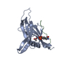
Mass: 60.052 Da / Num. of mol.: 1 / Source method: obtained synthetically / Formula: C2H4O2
#4: Water ChemComp-HOH / water


Mass: 18.015 Da / Num. of mol.: 220 / Source method: isolated from a natural source / Formula: H2O

-
Experimental details

-
Experiment

ExperimentMethod: X-RAY DIFFRACTION / Number of used crystals: 1

-
Sample preparation

CrystalDensity Matthews: 2.57 Å3/Da / Density % sol: 52.17 %
Crystal growTemperature: 298 K / Method: vapor diffusion, hanging drop / pH: 6
Details: PEG4K, PEG200, potassium acetate, pH 6.0, VAPOR DIFFUSION, HANGING DROP, temperature 298K

-
Data collection

DiffractionMean temperature: 100 K
Diffraction sourceSource: SYNCHROTRON / Site: CHESS / Beamline: F1
DetectorType: ADSC QUANTUM 4 / Detector: CCD / Date: Aug 1, 2004
RadiationProtocol: SINGLE WAVELENGTH / Monochromatic (M) / Laue (L): M / Scattering type: x-ray
Radiation wavelengthRelative weight: 1
ReflectionResolution: 1.7→100 Å / Num. all: 15581 / Num. obs: 15254 / % possible obs: 97.9 % / Biso Wilson estimate: 9.5 Å2

-
Processing

Software
NameVersionClassification
CNS1.1refinement
DENZOdata reduction
SCALEPACKdata scaling
CNSphasing
RefinementMethod to determine structure: MOLECULAR REPLACEMENT / Resolution: 1.7→34.35 Å / Rfactor Rfree error: 0.007 / Data cutoff high absF: 276148.03 / Data cutoff low absF: 0 / Isotropic thermal model: RESTRAINED / Cross valid method: THROUGHOUT / σ(F): 0
RfactorNum. reflection% reflectionSelection details
Rfree0.221 1043 6.8 %RANDOM
Rwork0.176 ---
obs0.176 15254 96.2 %-
all-15254 --
Solvent computationSolvent model: FLAT MODEL / Bsol: 41.0499 Å2 / ksol: 0.364346 e/Å3
Displacement parametersBiso mean: 12.9 Å2
Baniso -1Baniso -2Baniso -3
1-0.1 Å20 Å20 Å2
2--0.6 Å20 Å2
3----0.7 Å2
Refine analyze
FreeObs
Luzzati coordinate error0.21 Å0.16 Å
Luzzati d res low-5 Å
Luzzati sigma a0.09 Å0.05 Å
Refinement stepCycle: LAST / Resolution: 1.7→34.35 Å
ProteinNucleic acidLigandSolventTotal
Num. atoms1117 0 0 224 1341
Refine LS restraints
Refine-IDTypeDev idealDev ideal target
X-RAY DIFFRACTIONc_bond_d0.01
X-RAY DIFFRACTIONc_angle_deg1.5
X-RAY DIFFRACTIONc_dihedral_angle_d25.5
X-RAY DIFFRACTIONc_improper_angle_d0.95
X-RAY DIFFRACTIONc_mcbond_it1.241.5
X-RAY DIFFRACTIONc_mcangle_it1.652
X-RAY DIFFRACTIONc_scbond_it1.892
X-RAY DIFFRACTIONc_scangle_it2.792.5
LS refinement shellResolution: 1.7→1.81 Å / Rfactor Rfree error: 0.023 / Total num. of bins used: 6
RfactorNum. reflection% reflection
Rfree0.253 121 5.1 %
Rwork0.183 2257 -
obs--92.4 %
Xplor file
Refine-IDSerial noParam fileTopol file
X-RAY DIFFRACTION1PRO.PARAMPRO.TOP
X-RAY DIFFRACTION2WATER_REP.PARAM
X-RAY DIFFRACTION3ACY.PARAM

+
About Yorodumi

-
News

-
Feb 9, 2022. New format data for meta-information of EMDB entries

New format data for meta-information of EMDB entries

  • Version 3 of the EMDB header file is now the official format.
  • The previous official version 1.9 will be removed from the archive.

Related info.:EMDB header

External links:wwPDB to switch to version 3 of the EMDB data model

-
Aug 12, 2020. Covid-19 info

Covid-19 info

URL: https://pdbjlvh1.pdbj.org/emnavi/covid19.php

New page: Covid-19 featured information page in EM Navigator.

Related info.:Covid-19 info / Mar 5, 2020. Novel coronavirus structure data

+
Mar 5, 2020. Novel coronavirus structure data

Novel coronavirus structure data

Related info.:Yorodumi Speices / Aug 12, 2020. Covid-19 info

External links:COVID-19 featured content - PDBj / Molecule of the Month (242):Coronavirus Proteases

+
Jan 31, 2019. EMDB accession codes are about to change! (news from PDBe EMDB page)

EMDB accession codes are about to change! (news from PDBe EMDB page)

  • The allocation of 4 digits for EMDB accession codes will soon come to an end. Whilst these codes will remain in use, new EMDB accession codes will include an additional digit and will expand incrementally as the available range of codes is exhausted. The current 4-digit format prefixed with “EMD-” (i.e. EMD-XXXX) will advance to a 5-digit format (i.e. EMD-XXXXX), and so on. It is currently estimated that the 4-digit codes will be depleted around Spring 2019, at which point the 5-digit format will come into force.
  • The EM Navigator/Yorodumi systems omit the EMD- prefix.

Related info.:Q: What is EMD? / ID/Accession-code notation in Yorodumi/EM Navigator

External links:EMDB Accession Codes are Changing Soon! / Contact to PDBj

+
Jul 12, 2017. Major update of PDB

Major update of PDB

  • wwPDB released updated PDB data conforming to the new PDBx/mmCIF dictionary.
  • This is a major update changing the version number from 4 to 5, and with Remediation, in which all the entries are updated.
  • In this update, many items about electron microscopy experimental information are reorganized (e.g. em_software).
  • Now, EM Navigator and Yorodumi are based on the updated data.

External links:wwPDB Remediation / Enriched Model Files Conforming to OneDep Data Standards Now Available in the PDB FTP Archive

-
Yorodumi

Thousand views of thousand structures

  • Yorodumi is a browser for structure data from EMDB, PDB, SASBDB, etc.
  • This page is also the successor to EM Navigator detail page, and also detail information page/front-end page for Omokage search.
  • The word "yorodu" (or yorozu) is an old Japanese word meaning "ten thousand". "mi" (miru) is to see.

Related info.:EMDB / PDB / SASBDB / Comparison of 3 databanks / Yorodumi Search / Aug 31, 2016. New EM Navigator & Yorodumi / Yorodumi Papers / Jmol/JSmol / Function and homology information / Changes in new EM Navigator and Yorodumi

Read more