[English] 日本語
Yorodumi
- PDB-2pqu: Crystal structure of KH1 domain of human PCBP2 complexed to singl... -

+
Open data


ID or keywords:

Loading...

-
Basic information

Entry
Database: PDB / ID: 2pqu
TitleCrystal structure of KH1 domain of human PCBP2 complexed to single-stranded 12-mer telomeric dna
Components
  • 12-mer C-rich strand of human telomeric DNA
  • Poly(rC)-binding protein 2
KeywordsRNA AND DNA Binding protein/DNA / DNA Binding Protein-DNA complex / RNA AND DNA Binding protein-DNA COMPLEX
Function / homology
Function and homology information


mRNA metabolic process / regulation of RNA metabolic process / negative regulation of defense response to virus / IRES-dependent viral translational initiation / negative regulation of cGAS/STING signaling pathway / Processing of Capped Intron-Containing Pre-mRNA / mRNA Splicing - Major Pathway / Negative regulators of DDX58/IFIH1 signaling / SARS-CoV-1 activates/modulates innate immune responses / single-stranded DNA binding ...mRNA metabolic process / regulation of RNA metabolic process / negative regulation of defense response to virus / IRES-dependent viral translational initiation / negative regulation of cGAS/STING signaling pathway / Processing of Capped Intron-Containing Pre-mRNA / mRNA Splicing - Major Pathway / Negative regulators of DDX58/IFIH1 signaling / SARS-CoV-1 activates/modulates innate immune responses / single-stranded DNA binding / regulation of gene expression / proteasome-mediated ubiquitin-dependent protein catabolic process / defense response to virus / postsynaptic density / ribonucleoprotein complex / viral RNA genome replication / innate immune response / focal adhesion / mRNA binding / ubiquitin protein ligase binding / enzyme binding / RNA binding / extracellular exosome / nucleoplasm / membrane / nucleus / cytoplasm / cytosol
Similarity search - Function
K Homology domain, type 1 / KH domain / K Homology domain, type 1 / Ribosomal Protein S8; Chain: A, domain 1 / Type-1 KH domain profile. / K Homology domain, type 1 superfamily / K Homology domain / K homology RNA-binding domain / 2-Layer Sandwich / Alpha Beta
Similarity search - Domain/homology
DNA / DNA (> 10) / Poly(rC)-binding protein 2
Similarity search - Component
Biological speciesHomo sapiens (human)
MethodX-RAY DIFFRACTION / SYNCHROTRON / SAD / Resolution: 2.12 Å
AuthorsJames, T.L. / Lee, J.
CitationJournal: Rna / Year: 2007
Title: X-ray crystallographic and NMR studies of protein-protein and protein-nucleic acid interactions involving the KH domains from human poly(C)-binding protein-2.
Authors: Du, Z. / Lee, J.K. / Fenn, S. / Tjhen, R. / Stroud, R.M. / James, T.L.
History
DepositionMay 2, 2007Deposition site: RCSB / Processing site: RCSB
Revision 1.0Jun 12, 2007Provider: repository / Type: Initial release
Revision 1.1May 1, 2008Group: Version format compliance
Revision 1.2Jul 13, 2011Group: Advisory / Refinement description / Version format compliance
Revision 1.3Oct 18, 2017Group: Refinement description / Category: software
Item: _software.classification / _software.contact_author ..._software.classification / _software.contact_author / _software.contact_author_email / _software.date / _software.language / _software.location / _software.name / _software.type / _software.version
Revision 1.4Jul 24, 2019Group: Data collection / Derived calculations / Refinement description
Category: software / struct_conn
Item: _software.classification / _software.contact_author ..._software.classification / _software.contact_author / _software.contact_author_email / _software.date / _software.language / _software.location / _software.name / _software.type / _software.version / _struct_conn.pdbx_leaving_atom_flag

-
Structure visualization

Structure viewerMolecule:
MolmilJmol/JSmol

Downloads & links

-
Assembly

Deposited unit
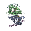
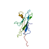
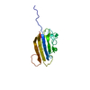
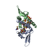
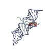
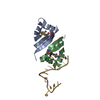
E: 12-mer C-rich strand of human telomeric DNA
G: 12-mer C-rich strand of human telomeric DNA
A: Poly(rC)-binding protein 2
B: Poly(rC)-binding protein 2
C: Poly(rC)-binding protein 2
D: Poly(rC)-binding protein 2


Theoretical massNumber of molelcules
Total (without water)39,7676
Polymers39,7676
Non-polymers00
Water2,396133
1
G: 12-mer C-rich strand of human telomeric DNA
A: Poly(rC)-binding protein 2
B: Poly(rC)-binding protein 2


Theoretical massNumber of molelcules
Total (without water)19,8843
Polymers19,8843
Non-polymers00
Water543
TypeNameSymmetry operationNumber
identity operation1_555x,y,z1
2
E: 12-mer C-rich strand of human telomeric DNA
C: Poly(rC)-binding protein 2
D: Poly(rC)-binding protein 2


Theoretical massNumber of molelcules
Total (without water)19,8843
Polymers19,8843
Non-polymers00
Water543
TypeNameSymmetry operationNumber
identity operation1_555x,y,z1
Unit cell
Length a, b, c (Å)92.228, 58.606, 71.738
Angle α, β, γ (deg.)90.00, 90.00, 90.00
Int Tables number18
Space group name H-MP21212
Noncrystallographic symmetry (NCS)NCS domain:
IDEns-IDDetails
11A
21B

NCS domain segments:

Component-ID: 1 / Ens-ID: 1 / Beg label comp-ID: VAL / End label comp-ID: LEU / Refine code: 4 / Auth seq-ID: 12 - 79 / Label seq-ID: 3 - 70

Dom-IDAuth asym-IDLabel asym-ID
1AC
2BD
DetailsThe biological assembly is a dimer of KH1 domains. One biological dimer is present in the asymmetric unit. Two domains are monomers in the asymmetric unit and the biological assembly is generated by the two fold axis.

-
Components

#1: DNA chain 12-mer C-rich strand of human telomeric DNA


Mass: 3551.346 Da / Num. of mol.: 2 / Source method: obtained synthetically
#2: Protein
Poly(rC)-binding protein 2 / Alpha-CP2 / hnRNP-E2


Mass: 8166.099 Da / Num. of mol.: 4 / Fragment: First KH domain of Human Poly(C)-Binding Protein
Source method: isolated from a genetically manipulated source
Source: (gene. exp.) Homo sapiens (human) / Gene: PCBP2 / Plasmid: pet24a / Species (production host): Escherichia coli / Production host: Escherichia coli BL21(DE3) (bacteria) / Strain (production host): BL21(DE3) / References: UniProt: Q15366
#3: Water ChemComp-HOH / water


Mass: 18.015 Da / Num. of mol.: 133 / Source method: isolated from a natural source / Formula: H2O

-
Experimental details

-
Experiment

ExperimentMethod: X-RAY DIFFRACTION / Number of used crystals: 1

-
Sample preparation

CrystalDensity Matthews: 2.45 Å3/Da / Density % sol: 49.71 %
Crystal growTemperature: 295 K / Method: vapor diffusion, hanging drop / pH: 6.1
Details: 25% PEG 8000, 100 mM sodium acetate, 100 mM sodium cacodylate , pH 6.1, VAPOR DIFFUSION, HANGING DROP, temperature 295K
Components of the solutions
IDNameCrystal-IDSol-ID
1PEG 800011
2sodium acetate11
3sodium cacodylate11
4PEG 800012
5sodium acetate12
6sodium cacodylate12

-
Data collection

DiffractionMean temperature: 100 K
Diffraction sourceSource: SYNCHROTRON / Site: ALS / Beamline: 8.3.1 / Wavelength: 0.979594 Å
DetectorType: ADSC QUANTUM 210 / Detector: CCD / Date: Jun 28, 2005
RadiationMonochromator: double crystal Si(111) / Protocol: SINGLE WAVELENGTH / Monochromatic (M) / Laue (L): M / Scattering type: x-ray
Radiation wavelengthWavelength: 0.979594 Å / Relative weight: 1
ReflectionRedundancy: 6.2 % / Av σ(I) over netI: 13.9 / Number: 201168 / Rmerge(I) obs: 0.11 / Χ2: 1.05 / D res high: 2.25 Å / D res low: 60 Å / Num. obs: 32413 / % possible obs: 89.8
Diffraction reflection shell
Highest resolution (Å)Lowest resolution (Å)% possible obs (%)IDRmerge(I) obsChi squaredRedundancy
5.156095.510.0411.0626.6
4.095.1598.610.0611.0566.6
3.574.0999.210.0841.0486.6
3.243.5799.410.1131.0186.6
3.013.2499.510.1440.9916.5
2.833.0199.610.1721.0596.3
2.692.8399.610.2331.0916
2.582.6999.610.2971.0895.8
2.482.5894.410.3381.1185.8
2.392.4877.410.3421.0695.6
2.322.3962.110.3941.0785.8
2.252.3253.210.4220.9735.7
ReflectionResolution: 2.1→50 Å / Num. obs: 22655 / % possible obs: 96.4 % / Observed criterion σ(F): 2 / Redundancy: 4.6 % / Rmerge(I) obs: 0.043 / Net I/σ(I): 18.5
Reflection shellResolution: 2.1→2.18 Å / Redundancy: 3.3 % / Rmerge(I) obs: 0.413 / Mean I/σ(I) obs: 2.72 / % possible all: 66.3

-
Phasing

PhasingMethod: SAD
Phasing dm shellResolution: 3.5→60 Å / Delta phi final: 0.16 / FOM : 0.173 / Reflection: 9254

-
Processing

Software
NameVersionClassificationNB
REFMACrefmac_5.2.0019refinement
SCALEPACKdata scaling
CNSphasing
CNSrefinement
DENZOdata reduction
PDB_EXTRACT2data extraction
Blu-Icedata collection
HKL-2000data reduction
RefinementMethod to determine structure: SAD / Resolution: 2.12→40 Å / Cor.coef. Fo:Fc: 0.948 / Cor.coef. Fo:Fc free: 0.929 / SU B: 13.922 / SU ML: 0.18 / TLS residual ADP flag: LIKELY RESIDUAL / Cross valid method: THROUGHOUT / ESU R: 0.239 / ESU R Free: 0.208 / Stereochemistry target values: MAXIMUM LIKELIHOOD / Details: HYDROGENS HAVE BEEN ADDED IN THE RIDING POSITIONS
RfactorNum. reflection% reflectionSelection details
Rfree0.26792 1156 5.1 %RANDOM
Rwork0.21506 ---
obs0.21772 21432 99.41 %-
Solvent computationIon probe radii: 0.8 Å / Shrinkage radii: 0.8 Å / VDW probe radii: 1.2 Å / Solvent model: MASK
Displacement parametersBiso mean: 43.317 Å2
Baniso -1Baniso -2Baniso -3
1--0.96 Å20 Å20 Å2
2---0.35 Å20 Å2
3---1.31 Å2
Refine analyzeLuzzati coordinate error obs: 0.44 Å / Luzzati d res low obs: 5 Å / Luzzati sigma a obs: 0.32 Å
Refinement stepCycle: LAST / Resolution: 2.12→40 Å
ProteinNucleic acidLigandSolventTotal
Num. atoms2149 470 0 133 2752
Refine LS restraints
Refine-IDTypeDev idealDev ideal targetNumber
X-RAY DIFFRACTIONr_bond_refined_d0.0240.0222693
X-RAY DIFFRACTIONr_bond_other_d
X-RAY DIFFRACTIONr_angle_refined_deg2.2482.2183694
X-RAY DIFFRACTIONr_angle_other_deg
X-RAY DIFFRACTIONr_dihedral_angle_1_deg7.6795278
X-RAY DIFFRACTIONr_dihedral_angle_2_deg43.77224.28677
X-RAY DIFFRACTIONr_dihedral_angle_3_deg18.97915461
X-RAY DIFFRACTIONr_dihedral_angle_4_deg26.5891516
X-RAY DIFFRACTIONr_chiral_restr0.1380.2436
X-RAY DIFFRACTIONr_gen_planes_refined0.0090.021752
X-RAY DIFFRACTIONr_gen_planes_other
X-RAY DIFFRACTIONr_nbd_refined0.2230.21108
X-RAY DIFFRACTIONr_nbd_other
X-RAY DIFFRACTIONr_nbtor_refined0.3010.21766
X-RAY DIFFRACTIONr_nbtor_other
X-RAY DIFFRACTIONr_xyhbond_nbd_refined0.2130.2111
X-RAY DIFFRACTIONr_xyhbond_nbd_other
X-RAY DIFFRACTIONr_metal_ion_refined
X-RAY DIFFRACTIONr_metal_ion_other
X-RAY DIFFRACTIONr_symmetry_vdw_refined0.3130.267
X-RAY DIFFRACTIONr_symmetry_vdw_other
X-RAY DIFFRACTIONr_symmetry_hbond_refined0.2290.215
X-RAY DIFFRACTIONr_symmetry_hbond_other
X-RAY DIFFRACTIONr_symmetry_metal_ion_refined
X-RAY DIFFRACTIONr_symmetry_metal_ion_other
X-RAY DIFFRACTIONr_mcbond_it2.11921456
X-RAY DIFFRACTIONr_mcbond_other
X-RAY DIFFRACTIONr_mcangle_it3.05232240
X-RAY DIFFRACTIONr_scbond_it1.95421509
X-RAY DIFFRACTIONr_scangle_it2.74631454
X-RAY DIFFRACTIONr_rigid_bond_restr
X-RAY DIFFRACTIONr_sphericity_free
X-RAY DIFFRACTIONr_sphericity_bonded
Refine LS restraints NCS

Dom-ID: 1 / Auth asym-ID: A / Ens-ID: 1 / Number: 507 / Refine-ID: X-RAY DIFFRACTION

TypeRms dev position (Å)Weight position
MEDIUM POSITIONAL0.80.5
MEDIUM THERMAL2.242
LS refinement shellResolution: 2.12→2.175 Å / Total num. of bins used: 20
RfactorNum. reflection% reflection
Rfree0.348 82 -
Rwork0.287 1516 -
obs--95.98 %
Refinement TLS params.

Method: refined / Refine-ID: X-RAY DIFFRACTION

IDL112)L122)L132)L222)L232)L332)S11 (Å °)S12 (Å °)S13 (Å °)S21 (Å °)S22 (Å °)S23 (Å °)S31 (Å °)S32 (Å °)S33 (Å °)T112)T122)T132)T222)T232)T332)Origin x (Å)Origin y (Å)Origin z (Å)
16.07630.33190.30863.6643-1.72195.68570.1590.5954-1.03210.0787-0.2434-0.0826-0.0484-0.02750.0844-0.16470.0387-0.0455-0.1924-0.1840.070650.6116.9229.929
27.1428-1.27910.10932.91140.48694.86280.149-0.5712-1.2926-0.06080.07960.3619-0.0174-0.1008-0.2285-0.1314-0.01970.0169-0.21320.12660.067944.62917.09426.873
37.25570.3271-1.16352.56790.15782.6775-0.0261-0.4320.34320.0157-0.1512-0.0831-0.19730.41850.1773-0.12110.0118-0.00340.04010.0423-0.141529.75110.392-15.807
49.5603-4.91997.09652.9488-3.74665.4853-0.1213-0.17360.9873-0.14150.3254-0.62450.27280.2273-0.2041-0.1015-0.0395-0.04270.0751-0.16950.115842.42916.141-9.482
58.8041.54436.72240.83280.49965.9549-0.22280.13051.09390.25720.15910.352-0.0236-0.29120.0637-0.05810.10170.027-0.02710.15020.140452.9415.11545.753
Refinement TLS group
IDRefine-IDRefine TLS-IDAuth asym-IDAuth seq-ID
1X-RAY DIFFRACTION1A11 - 80
2X-RAY DIFFRACTION2B12 - 79
3X-RAY DIFFRACTION3C12 - 79
4X-RAY DIFFRACTION4E498 - 509
5X-RAY DIFFRACTION5G499 - 510

+
About Yorodumi

-
News

-
Feb 9, 2022. New format data for meta-information of EMDB entries

New format data for meta-information of EMDB entries

  • Version 3 of the EMDB header file is now the official format.
  • The previous official version 1.9 will be removed from the archive.

Related info.:EMDB header

External links:wwPDB to switch to version 3 of the EMDB data model

-
Aug 12, 2020. Covid-19 info

Covid-19 info

URL: https://pdbjlvh1.pdbj.org/emnavi/covid19.php

New page: Covid-19 featured information page in EM Navigator.

Related info.:Covid-19 info / Mar 5, 2020. Novel coronavirus structure data

+
Mar 5, 2020. Novel coronavirus structure data

Novel coronavirus structure data

Related info.:Yorodumi Speices / Aug 12, 2020. Covid-19 info

External links:COVID-19 featured content - PDBj / Molecule of the Month (242):Coronavirus Proteases

+
Jan 31, 2019. EMDB accession codes are about to change! (news from PDBe EMDB page)

EMDB accession codes are about to change! (news from PDBe EMDB page)

  • The allocation of 4 digits for EMDB accession codes will soon come to an end. Whilst these codes will remain in use, new EMDB accession codes will include an additional digit and will expand incrementally as the available range of codes is exhausted. The current 4-digit format prefixed with “EMD-” (i.e. EMD-XXXX) will advance to a 5-digit format (i.e. EMD-XXXXX), and so on. It is currently estimated that the 4-digit codes will be depleted around Spring 2019, at which point the 5-digit format will come into force.
  • The EM Navigator/Yorodumi systems omit the EMD- prefix.

Related info.:Q: What is EMD? / ID/Accession-code notation in Yorodumi/EM Navigator

External links:EMDB Accession Codes are Changing Soon! / Contact to PDBj

+
Jul 12, 2017. Major update of PDB

Major update of PDB

  • wwPDB released updated PDB data conforming to the new PDBx/mmCIF dictionary.
  • This is a major update changing the version number from 4 to 5, and with Remediation, in which all the entries are updated.
  • In this update, many items about electron microscopy experimental information are reorganized (e.g. em_software).
  • Now, EM Navigator and Yorodumi are based on the updated data.

External links:wwPDB Remediation / Enriched Model Files Conforming to OneDep Data Standards Now Available in the PDB FTP Archive

-
Yorodumi

Thousand views of thousand structures

  • Yorodumi is a browser for structure data from EMDB, PDB, SASBDB, etc.
  • This page is also the successor to EM Navigator detail page, and also detail information page/front-end page for Omokage search.
  • The word "yorodu" (or yorozu) is an old Japanese word meaning "ten thousand". "mi" (miru) is to see.

Related info.:EMDB / PDB / SASBDB / Comparison of 3 databanks / Yorodumi Search / Aug 31, 2016. New EM Navigator & Yorodumi / Yorodumi Papers / Jmol/JSmol / Function and homology information / Changes in new EM Navigator and Yorodumi

Read more