 Movie
Movie Controller

[English] 日本語
 Yorodumi
- PDB-5hr7: X-ray crystal structure of C118A RlmN from Escherichia coli with ... -
+ Open data

- Basic information
| Entry | Database: PDB / ID: 5hr7 | ||||||
|---|---|---|---|---|---|---|---|
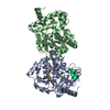
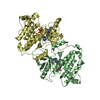
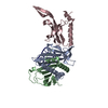
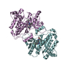
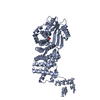
| Title | X-ray crystal structure of C118A RlmN from Escherichia coli with cross-linked in vitro transcribed tRNA | ||||||
 Components Components |
| ||||||
 Keywords Keywords | OXIDOREDUCTASE/RNA / protein-RNA complex / radical SAM enzyme / transfer RNA / iron-sulfur cluster / OXIDOREDUCTASE-RNA complex | ||||||
| Function / homology |  Function and homology information Function and homology information23S rRNA (adenine2503-C2)-methyltransferase / tRNA (adenine(37)-C2)-methyltransferase activity / rRNA (adenine(2503)-C2-)-methyltransferase activity / tRNA methylation / rRNA base methylation / 4 iron, 4 sulfur cluster binding / tRNA binding / rRNA binding / response to antibiotic / metal ion binding ...23S rRNA (adenine2503-C2)-methyltransferase / tRNA (adenine(37)-C2)-methyltransferase activity / rRNA (adenine(2503)-C2-)-methyltransferase activity / tRNA methylation / rRNA base methylation / 4 iron, 4 sulfur cluster binding / tRNA binding / rRNA binding / response to antibiotic / metal ion binding / cytosol / cytoplasm Similarity search - Function | ||||||
| Biological species |  | ||||||
| Method |  X-RAY DIFFRACTION / SYNCHROTRON / MOLECULAR REPLACEMENT / Resolution: 2.4 Å X-RAY DIFFRACTION / SYNCHROTRON / MOLECULAR REPLACEMENT / Resolution: 2.4 Å | ||||||
 Authors Authors | Schwalm, E.L. / Grove, T.L. / Booker, S.J. / Boal, A.K. | ||||||
 Citation Citation | Journal: Science / Year: 2016 Title: Crystallographic capture of a radical S-adenosylmethionine enzyme in the act of modifying tRNA. Authors: Schwalm, E.L. / Grove, T.L. / Booker, S.J. / Boal, A.K. | ||||||
| History |
|
- Structure visualization
| Structure viewer | Molecule: MolmilJmol/JSmol |
|---|
- Downloads & links
-Download
| PDBx/mmCIF format | 5hr7.cif.gz | 237.6 KB | Display | PDBx/mmCIF format |
|---|---|---|---|---|
| PDB format | pdb5hr7.ent.gz | 182.8 KB | Display | PDB format |
| PDBx/mmJSON format | 5hr7.json.gz | Tree view | PDBx/mmJSON format | |
| Others |  Other downloads Other downloads |
-Validation report
| Arichive directory | https://data.pdbj.org/pub/pdb/validation_reports/hr/5hr7ftp://data.pdbj.org/pub/pdb/validation_reports/hr/5hr7 | HTTPS FTP |
|---|
-Related structure data
| Related structure data |  5hr6C  2derS  3rfaS C: citing same article ( S: Starting model for refinement |
|---|---|
| Similar structure data |


-Links
 PDBj
PDBj


- Assembly
| Deposited unit | 
| ||||||||
|---|---|---|---|---|---|---|---|---|---|
| 1 | 
| ||||||||
| 2 | 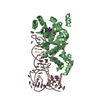
| ||||||||
| Unit cell |
|
-Components
-RNA chain / Protein , 2 types, 4 molecules DCBA
| #1: RNA chain | Mass: 24378.514 Da / Num. of mol.: 2 / Source method: obtained synthetically Details: RNA was prepared by in vitro transcription with T7 RNA polymerase Source: (synth.) #2: Protein | Mass: 45400.766 Da / Num. of mol.: 2 Source method: isolated from a genetically manipulated source Source: (gene. exp.) References: UniProt: A7ZPW0, UniProt: P36979*PLUS, 23S rRNA (adenine2503-C2)-methyltransferase |
|---|
-Non-polymers , 5 types, 107 molecules 


| #3: Chemical | ChemComp-MG /  Mass: 24.305 Da / Num. of mol.: 7 / Source method: obtained synthetically / Formula: Mg Mass: 24.305 Da / Num. of mol.: 7 / Source method: obtained synthetically / Formula: Mg#4: Chemical |  Mass: 351.640 Da / Num. of mol.: 2 / Source method: obtained synthetically / Formula: Fe4S4 Mass: 351.640 Da / Num. of mol.: 2 / Source method: obtained synthetically / Formula: Fe4S4#5: Chemical |  Mass: 251.242 Da / Num. of mol.: 2 / Source method: obtained synthetically / Formula: C10H13N5O3 Mass: 251.242 Da / Num. of mol.: 2 / Source method: obtained synthetically / Formula: C10H13N5O3#6: Chemical |  Type: L-peptide linking / Mass: 149.211 Da / Num. of mol.: 2 / Source method: obtained synthetically / Formula: C5H11NO2S Type: L-peptide linking / Mass: 149.211 Da / Num. of mol.: 2 / Source method: obtained synthetically / Formula: C5H11NO2S#7: Water | ChemComp-HOH / | Mass: 18.015 Da / Num. of mol.: 94 / Source method: isolated from a natural source / Formula: H2O |
|---|
-Details
| Has protein modification | Y |
|---|
-Experimental details
-Experiment
| Experiment | Method: X-RAY DIFFRACTION / Number of used crystals: 1 |
|---|
- Sample preparation
| Crystal | Density Matthews: 3.47 Å3/Da / Density % sol: 64.55 % |
|---|---|
| Crystal grow | Temperature: 298 K / Method: vapor diffusion / pH: 6 Details: PEG 4000, sodium chloride, sodium cacodylate trihydrate, spermine |
-Data collection
| Diffraction | Mean temperature: 100 K |
|---|---|
| Diffraction source | Source: SYNCHROTRON / Site: APS  / Beamline: 21-ID-G / Wavelength: 0.9786 Å / Beamline: 21-ID-G / Wavelength: 0.9786 Å |
| Detector | Type: MARMOSAIC 300 mm CCD / Detector: CCD / Date: Oct 26, 2014 |
| Radiation | Protocol: SINGLE WAVELENGTH / Monochromatic (M) / Laue (L): M / Scattering type: x-ray |
| Radiation wavelength | Wavelength: 0.9786 Å / Relative weight: 1 |
| Reflection | Resolution: 2.4→50 Å / Num. obs: 75380 / % possible obs: 99.9 % / Redundancy: 7.3 % / CC1/2: 0.8 / Rmerge(I) obs: 0.087 / Net I/σ(I): 34.9 |
| Reflection shell | Resolution: 2.4→2.44 Å / Redundancy: 5.7 % / Rmerge(I) obs: 0.529 / Mean I/σ(I) obs: 1.8 / % possible all: 99.2 |
- Processing
| Software |
| |||||||||||||||||||||||||||||||||||||||||||||||||||||||||||||||||||||||||||
|---|---|---|---|---|---|---|---|---|---|---|---|---|---|---|---|---|---|---|---|---|---|---|---|---|---|---|---|---|---|---|---|---|---|---|---|---|---|---|---|---|---|---|---|---|---|---|---|---|---|---|---|---|---|---|---|---|---|---|---|---|---|---|---|---|---|---|---|---|---|---|---|---|---|---|---|---|
| Refinement | Method to determine structure: MOLECULAR REPLACEMENT Starting model: 3RFA,2DER Resolution: 2.4→50 Å / Cor.coef. Fo:Fc: 0.942 / Cor.coef. Fo:Fc free: 0.929 / SU B: 7.669 / SU ML: 0.175 / Cross valid method: THROUGHOUT / σ(F): 0 / ESU R: 0.26 / ESU R Free: 0.212 Details: HYDROGENS HAVE BEEN ADDED IN THE RIDING POSITIONS U VALUES : REFINED INDIVIDUALLY
| |||||||||||||||||||||||||||||||||||||||||||||||||||||||||||||||||||||||||||
| Solvent computation | Ion probe radii: 0.8 Å / Shrinkage radii: 0.8 Å / VDW probe radii: 1.2 Å | |||||||||||||||||||||||||||||||||||||||||||||||||||||||||||||||||||||||||||
| Displacement parameters | Biso max: 141.32 Å2 / Biso mean: 62.237 Å2 / Biso min: 35.79 Å2
| |||||||||||||||||||||||||||||||||||||||||||||||||||||||||||||||||||||||||||
| Refinement step | Cycle: final / Resolution: 2.4→50 Å
| |||||||||||||||||||||||||||||||||||||||||||||||||||||||||||||||||||||||||||
| Refine LS restraints |
| |||||||||||||||||||||||||||||||||||||||||||||||||||||||||||||||||||||||||||
| LS refinement shell | Resolution: 2.397→2.459 Å / Total num. of bins used: 20
|
