[English] 日本語
Yorodumi
- PDB-5hq8: Co-crystal Structure of human SMYD3 with a MEKK2 peptide at 2.13A -

+
Open data


ID or keywords:

Loading...

-
Basic information

Entry
Database: PDB / ID: 5hq8
TitleCo-crystal Structure of human SMYD3 with a MEKK2 peptide at 2.13A
Components
  • Histone-lysine N-methyltransferase SMYD3
  • MEKK2 peptide
KeywordsTRANSFERASE / SMYD3 / methyltransferase / oncology
Function / homology
Function and homology information


histone H3K36 dimethyltransferase activity / histone H4 methyltransferase activity / [histone H3]-lysine4 N-trimethyltransferase / myotube cell development / mitogen-activated protein kinase kinase kinase / histone H3K4 trimethyltransferase activity / RNA polymerase II intronic transcription regulatory region sequence-specific DNA binding / RNA polymerase II complex binding / MAP kinase kinase kinase activity / cellular response to dexamethasone stimulus ...histone H3K36 dimethyltransferase activity / histone H4 methyltransferase activity / [histone H3]-lysine4 N-trimethyltransferase / myotube cell development / mitogen-activated protein kinase kinase kinase / histone H3K4 trimethyltransferase activity / RNA polymerase II intronic transcription regulatory region sequence-specific DNA binding / RNA polymerase II complex binding / MAP kinase kinase kinase activity / cellular response to dexamethasone stimulus / establishment of protein localization / PKMTs methylate histone lysines / cellular response to mechanical stimulus / nucleosome assembly / positive regulation of peptidyl-serine phosphorylation / methylation / protein kinase activity / intracellular signal transduction / RNA polymerase II cis-regulatory region sequence-specific DNA binding / phosphorylation / protein serine kinase activity / protein serine/threonine kinase activity / protein kinase binding / positive regulation of transcription by RNA polymerase II / nucleoplasm / ATP binding / nucleus / metal ion binding / cytoplasm / cytosol
Similarity search - Function
Mitogen-activated protein kinase kinase kinase 2/3, PB1 domain / Annexin V; domain 1 - #160 / Serine Threonine Protein Phosphatase 5, Tetratricopeptide repeat - #970 / Helix Hairpins - #2220 / Histone-lysine N-methyltransferase Smyd3 / SMYD3, SET domain / Histone-lysine N-methyltransferase (EC 2.1.1.43) family profile. / PB1 domain / PB1 domain / PB1 domain profile. ...Mitogen-activated protein kinase kinase kinase 2/3, PB1 domain / Annexin V; domain 1 - #160 / Serine Threonine Protein Phosphatase 5, Tetratricopeptide repeat - #970 / Helix Hairpins - #2220 / Histone-lysine N-methyltransferase Smyd3 / SMYD3, SET domain / Histone-lysine N-methyltransferase (EC 2.1.1.43) family profile. / PB1 domain / PB1 domain / PB1 domain profile. / PB1 domain / MYND finger / Zinc finger, MYND-type / Zinc finger MYND-type signature. / Zinc finger MYND-type profile. / Annexin V; domain 1 / Beta-clip-like / SET domain / Tetratricopeptide repeat domain / Helix Hairpins / SET (Su(var)3-9, Enhancer-of-zeste, Trithorax) domain / SET domain superfamily / SET domain / SET domain profile. / SET domain / Beta Complex / Helix non-globular / Serine Threonine Protein Phosphatase 5, Tetratricopeptide repeat / Special / Alpha Horseshoe / Tetratricopeptide-like helical domain superfamily / Protein kinase domain / Serine/Threonine protein kinases, catalytic domain / Protein kinase, ATP binding site / Protein kinases ATP-binding region signature. / Protein kinase domain profile. / Protein kinase domain / Protein kinase-like domain superfamily / Orthogonal Bundle / Mainly Beta / Mainly Alpha
Similarity search - Domain/homology
S-ADENOSYL-L-HOMOCYSTEINE / Histone-lysine N-methyltransferase SMYD3 / Mitogen-activated protein kinase kinase kinase 2
Similarity search - Component
Biological speciesHomo sapiens (human)
MethodX-RAY DIFFRACTION / SYNCHROTRON / MOLECULAR REPLACEMENT / Resolution: 1.72 Å
AuthorsElkins, P.A. / Bonnette, W.G.
CitationJournal: Structure / Year: 2016
Title: Structure-Based Design of a Novel SMYD3 Inhibitor that Bridges the SAM-and MEKK2-Binding Pockets.
Authors: Van Aller, G.S. / Graves, A.P. / Elkins, P.A. / Bonnette, W.G. / McDevitt, P.J. / Zappacosta, F. / Annan, R.S. / Dean, T.W. / Su, D.S. / Carpenter, C.L. / Mohammad, H.P. / Kruger, R.G.
History
DepositionJan 21, 2016Deposition site: RCSB / Processing site: RCSB
Revision 1.0Mar 30, 2016Provider: repository / Type: Initial release
Revision 1.1Apr 27, 2016Group: Database references
Revision 1.2May 18, 2016Group: Database references
Revision 1.3Mar 6, 2024Group: Data collection / Database references / Derived calculations
Category: chem_comp_atom / chem_comp_bond ...chem_comp_atom / chem_comp_bond / citation / database_2 / pdbx_struct_oper_list / struct_conn
Item: _citation.journal_id_CSD / _database_2.pdbx_DOI ..._citation.journal_id_CSD / _database_2.pdbx_DOI / _database_2.pdbx_database_accession / _pdbx_struct_oper_list.symmetry_operation / _struct_conn.pdbx_dist_value / _struct_conn.ptnr1_auth_asym_id / _struct_conn.ptnr1_auth_comp_id / _struct_conn.ptnr1_auth_seq_id / _struct_conn.ptnr1_label_asym_id / _struct_conn.ptnr1_label_atom_id / _struct_conn.ptnr1_label_comp_id / _struct_conn.ptnr1_label_seq_id / _struct_conn.ptnr2_auth_asym_id / _struct_conn.ptnr2_auth_comp_id / _struct_conn.ptnr2_auth_seq_id / _struct_conn.ptnr2_label_asym_id / _struct_conn.ptnr2_label_atom_id / _struct_conn.ptnr2_label_comp_id

-
Structure visualization

Structure viewerMolecule:
MolmilJmol/JSmol

Downloads & links

-
Assembly

Deposited unit
A: Histone-lysine N-methyltransferase SMYD3
B: Histone-lysine N-methyltransferase SMYD3
I: MEKK2 peptide
J: MEKK2 peptide
hetero molecules


Theoretical massNumber of molelcules
Total (without water)104,23121
Polymers102,5574
Non-polymers1,67417
Water12,917717
1
A: Histone-lysine N-methyltransferase SMYD3
I: MEKK2 peptide
hetero molecules


Theoretical massNumber of molelcules
Total (without water)52,07010
Polymers51,2782
Non-polymers7918
Water362
TypeNameSymmetry operationNumber
identity operation1_555x,y,z1
Buried area2610 Å2
ΔGint-7 kcal/mol
Surface area19320 Å2
MethodPISA
2
B: Histone-lysine N-methyltransferase SMYD3
J: MEKK2 peptide
hetero molecules


Theoretical massNumber of molelcules
Total (without water)52,16211
Polymers51,2782
Non-polymers8839
Water362
TypeNameSymmetry operationNumber
identity operation1_555x,y,z1
Buried area2680 Å2
ΔGint-2 kcal/mol
Surface area18640 Å2
MethodPISA
Unit cell
Length a, b, c (Å)53.109, 118.155, 84.782
Angle α, β, γ (deg.)90.00, 90.61, 90.00
Int Tables number4
Space group name H-MP1211

-
Components

-
Protein / Protein/peptide , 2 types, 4 molecules ABIJ

#1: Protein Histone-lysine N-methyltransferase SMYD3 / SET and MYND domain-containing protein 3 / Zinc finger MYND domain-containing protein 1


Mass: 49542.520 Da / Num. of mol.: 2
Source method: isolated from a genetically manipulated source
Source: (gene. exp.) Homo sapiens (human) / Gene: SMYD3, ZMYND1, ZNFN3A1 / Production host: unidentified baculovirus / Strain (production host): sf9
References: UniProt: Q9H7B4, histone-lysine N-methyltransferase
#2: Protein/peptide MEKK2 peptide


Mass: 1735.914 Da / Num. of mol.: 2 / Source method: obtained synthetically / Source: (synth.) Homo sapiens (human) / References: UniProt: Q9Y2U5*PLUS

-
Non-polymers , 6 types, 734 molecules

#3: Chemical
ChemComp-ZN / ZINC ION


Mass: 65.409 Da / Num. of mol.: 6 / Source method: obtained synthetically / Formula: Zn
#4: Chemical ChemComp-MG / MAGNESIUM ION


Mass: 24.305 Da / Num. of mol.: 2 / Source method: obtained synthetically / Formula: Mg
#5: Chemical
ChemComp-EDO / 1,2-ETHANEDIOL / ETHYLENE GLYCOL


Mass: 62.068 Da / Num. of mol.: 6 / Source method: obtained synthetically / Formula: C2H6O2
#6: Chemical ChemComp-SAH / S-ADENOSYL-L-HOMOCYSTEINE


Type: L-peptide linking / Mass: 384.411 Da / Num. of mol.: 2 / Source method: obtained synthetically / Formula: C14H20N6O5S
#7: Chemical ChemComp-GOL / GLYCEROL / GLYCERIN / PROPANE-1,2,3-TRIOL


Mass: 92.094 Da / Num. of mol.: 1 / Source method: obtained synthetically / Formula: C3H8O3
#8: Water ChemComp-HOH / water


Mass: 18.015 Da / Num. of mol.: 717 / Source method: isolated from a natural source / Formula: H2O

-
Experimental details

-
Experiment

ExperimentMethod: X-RAY DIFFRACTION / Number of used crystals: 1

-
Sample preparation

CrystalDensity Matthews: 2.63 Å3/Da / Density % sol: 53.28 %
Crystal grow
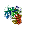
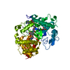
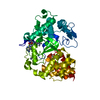
Temperature: 295 K / Method: vapor diffusion, sitting drop / pH: 7
Details: Human SMYD3 (1-428) with SAM in the SAM-binding site was concentrated to 11.7 mg/mL in 25 mM Tris-HCl pH 8 and 150 mM NaCl. Peptide (21st Century Biochemical) was solubilized in 100mM water. ...Details: Human SMYD3 (1-428) with SAM in the SAM-binding site was concentrated to 11.7 mg/mL in 25 mM Tris-HCl pH 8 and 150 mM NaCl. Peptide (21st Century Biochemical) was solubilized in 100mM water. Two ul peptide was added to 200 ul of protein (five fold molar excess of peptide) and allowed to complex with the protein overnight at 4C. Crystals were obtained from a Hampton Index HT broad screen in sitting drops at 22C with condition H7 (0.15 DL-malic acid pH 7.0, 20% w/v PEG 3,350). Crystals were flash frozen in 20 percent ethylene glycol in well solution prior to data collection

-
Data collection

DiffractionMean temperature: 100 K
Diffraction sourceSource: SYNCHROTRON / Site: APS / Beamline: 21-ID-D / Wavelength: 1.07808 Å
DetectorType: MARMOSAIC 300 mm CCD / Detector: CCD / Date: Nov 12, 2014
RadiationProtocol: SINGLE WAVELENGTH / Monochromatic (M) / Laue (L): M / Scattering type: x-ray
Radiation wavelengthWavelength: 1.07808 Å / Relative weight: 1
ReflectionResolution: 1.592→50 Å / Num. obs: 138107 / % possible obs: 99.8 % / Redundancy: 3.6 % / Net I/σ(I): 16.11

-
Processing

Software
NameVersionClassification
PHENIXdev_1801refinement
HKL-2000data reduction
HKL-2000data scaling
PHENIXphasing
RefinementMethod to determine structure: MOLECULAR REPLACEMENT / Resolution: 1.72→25.43 Å / SU ML: 0.19 / Cross valid method: FREE R-VALUE / σ(F): 1.36 / Phase error: 19.69 / Stereochemistry target values: ML
RfactorNum. reflection% reflection
Rfree0.2011 5551 5.03 %
Rwork0.1741 --
obs0.1755 110414 99.78 %
Solvent computationShrinkage radii: 0.9 Å / VDW probe radii: 1.11 Å / Solvent model: FLAT BULK SOLVENT MODEL
Refinement stepCycle: LAST / Resolution: 1.72→25.43 Å
ProteinNucleic acidLigandSolventTotal
Num. atoms6774 0 90 717 7581
Refine LS restraints
Refine-IDTypeDev idealNumber
X-RAY DIFFRACTIONf_bond_d0.0187104
X-RAY DIFFRACTIONf_angle_d1.4199587
X-RAY DIFFRACTIONf_dihedral_angle_d13.6232692
X-RAY DIFFRACTIONf_chiral_restr0.0911063
X-RAY DIFFRACTIONf_plane_restr0.0111234
LS refinement shell
Resolution (Å)Rfactor RfreeNum. reflection RfreeRfactor RworkNum. reflection RworkRefine-ID% reflection obs (%)
1.72-1.73950.28181920.24933473X-RAY DIFFRACTION100
1.7395-1.760.31860.22673524X-RAY DIFFRACTION100
1.76-1.78150.23741860.22283462X-RAY DIFFRACTION100
1.7815-1.8040.25991930.21673493X-RAY DIFFRACTION100
1.804-1.82770.23971820.21253500X-RAY DIFFRACTION100
1.8277-1.85280.24451800.20723469X-RAY DIFFRACTION100
1.8528-1.87920.23381930.20553505X-RAY DIFFRACTION100
1.8792-1.90730.22581880.20613488X-RAY DIFFRACTION100
1.9073-1.93710.22791800.19413466X-RAY DIFFRACTION100
1.9371-1.96880.24261830.19373542X-RAY DIFFRACTION100
1.9688-2.00270.22411840.19593442X-RAY DIFFRACTION100
2.0027-2.03910.20842060.20193496X-RAY DIFFRACTION100
2.0391-2.07830.21921790.18183495X-RAY DIFFRACTION100
2.0783-2.12080.21911790.18273539X-RAY DIFFRACTION100
2.1208-2.16680.23051790.18433445X-RAY DIFFRACTION100
2.1668-2.21720.20681950.17763510X-RAY DIFFRACTION100
2.2172-2.27260.19861930.17383490X-RAY DIFFRACTION100
2.2726-2.3340.21412030.17443459X-RAY DIFFRACTION100
2.334-2.40270.21081830.17833490X-RAY DIFFRACTION100
2.4027-2.48020.20781770.18193484X-RAY DIFFRACTION100
2.4802-2.56870.21031880.18223518X-RAY DIFFRACTION100
2.5687-2.67150.20711710.18623507X-RAY DIFFRACTION100
2.6715-2.79290.21041700.18163545X-RAY DIFFRACTION100
2.7929-2.93990.20841840.18273480X-RAY DIFFRACTION100
2.9399-3.12380.21441930.18573503X-RAY DIFFRACTION100
3.1238-3.36450.22171820.17653493X-RAY DIFFRACTION100
3.3645-3.70220.1781730.16413525X-RAY DIFFRACTION100
3.7022-4.23580.15881770.14093501X-RAY DIFFRACTION100
4.2358-5.32860.14811850.13613530X-RAY DIFFRACTION99
5.3286-25.43260.1841870.15313489X-RAY DIFFRACTION98
Refinement TLS params.

Method: refined / Refine-ID: X-RAY DIFFRACTION

IDL112)L122)L132)L222)L232)L332)S11 (Å °)S12 (Å °)S13 (Å °)S21 (Å °)S22 (Å °)S23 (Å °)S31 (Å °)S32 (Å °)S33 (Å °)T112)T122)T132)T222)T232)T332)Origin x (Å)Origin y (Å)Origin z (Å)
11.43840.65380.58892.05411.24072.56290.0013-0.06520.19770.02770.175-0.21420.02130.2445-0.1370.1011-0.0011-0.00060.1912-0.03010.241336.7776-5.99340.059
20.8928-0.46820.30082.54190.85783.00480.0552-0.09390.01230.06350.02760.1253-0.1333-0.0052-0.09550.0906-0.00650.00550.1695-0.05160.279223.5971-0.099943.6186
31.30540.16910.03651.368-0.16890.499-0.102-0.1027-0.16750.03940.0407-0.1643-0.00960.0470.05020.10380.00010.01750.1568-0.0070.157133.8138-27.694438.1049
Refinement TLS group
IDRefine-IDRefine TLS-IDSelection details
1X-RAY DIFFRACTION1chain 'A' and (resseq 3:93)
2X-RAY DIFFRACTION2chain 'A' and (resseq 94:186)
3X-RAY DIFFRACTION3chain 'A' and (resseq 187:427)

+
About Yorodumi

-
News

-
Feb 9, 2022. New format data for meta-information of EMDB entries

New format data for meta-information of EMDB entries

  • Version 3 of the EMDB header file is now the official format.
  • The previous official version 1.9 will be removed from the archive.

Related info.:EMDB header

External links:wwPDB to switch to version 3 of the EMDB data model

-
Aug 12, 2020. Covid-19 info

Covid-19 info

URL: https://pdbjlvh1.pdbj.org/emnavi/covid19.php

New page: Covid-19 featured information page in EM Navigator.

Related info.:Covid-19 info / Mar 5, 2020. Novel coronavirus structure data

+
Mar 5, 2020. Novel coronavirus structure data

Novel coronavirus structure data

Related info.:Yorodumi Speices / Aug 12, 2020. Covid-19 info

External links:COVID-19 featured content - PDBj / Molecule of the Month (242):Coronavirus Proteases

+
Jan 31, 2019. EMDB accession codes are about to change! (news from PDBe EMDB page)

EMDB accession codes are about to change! (news from PDBe EMDB page)

  • The allocation of 4 digits for EMDB accession codes will soon come to an end. Whilst these codes will remain in use, new EMDB accession codes will include an additional digit and will expand incrementally as the available range of codes is exhausted. The current 4-digit format prefixed with “EMD-” (i.e. EMD-XXXX) will advance to a 5-digit format (i.e. EMD-XXXXX), and so on. It is currently estimated that the 4-digit codes will be depleted around Spring 2019, at which point the 5-digit format will come into force.
  • The EM Navigator/Yorodumi systems omit the EMD- prefix.

Related info.:Q: What is EMD? / ID/Accession-code notation in Yorodumi/EM Navigator

External links:EMDB Accession Codes are Changing Soon! / Contact to PDBj

+
Jul 12, 2017. Major update of PDB

Major update of PDB

  • wwPDB released updated PDB data conforming to the new PDBx/mmCIF dictionary.
  • This is a major update changing the version number from 4 to 5, and with Remediation, in which all the entries are updated.
  • In this update, many items about electron microscopy experimental information are reorganized (e.g. em_software).
  • Now, EM Navigator and Yorodumi are based on the updated data.

External links:wwPDB Remediation / Enriched Model Files Conforming to OneDep Data Standards Now Available in the PDB FTP Archive

-
Yorodumi

Thousand views of thousand structures

  • Yorodumi is a browser for structure data from EMDB, PDB, SASBDB, etc.
  • This page is also the successor to EM Navigator detail page, and also detail information page/front-end page for Omokage search.
  • The word "yorodu" (or yorozu) is an old Japanese word meaning "ten thousand". "mi" (miru) is to see.

Related info.:EMDB / PDB / SASBDB / Comparison of 3 databanks / Yorodumi Search / Aug 31, 2016. New EM Navigator & Yorodumi / Yorodumi Papers / Jmol/JSmol / Function and homology information / Changes in new EM Navigator and Yorodumi

Read more