Mass: 18.015 Da / Num. of mol.: 119 / Source method: isolated from a natural source / Formula: H2O
-
Experimental details
-
Experiment
Experiment
Method: X-RAY DIFFRACTION / Number of used crystals: 1
-
Sample preparation
Crystal
Density Matthews: 2.12 Å3/Da / Density % sol: 42.1 %
Crystal grow
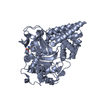
Temperature: 295 K / Method: vapor diffusion, sitting drop Details: SMYD3 crystallization and soaking of GSK2807: SMYD3 (1-428) expressed in baculovirus and with SAH in the SAM-binding site was crystallized in sitting drops at 22C using 1uL fresh protein at ...Details: SMYD3 crystallization and soaking of GSK2807: SMYD3 (1-428) expressed in baculovirus and with SAH in the SAM-binding site was crystallized in sitting drops at 22C using 1uL fresh protein at 8.9mg/mL with the addition of 1uL mother liquor (200mM MgOAc Tetrahydrate, 20% PEG 3350). Crystals grew to a large size but varied in quantity when set up in replicates. A seed stock was formed from these crystals and added at 20% to the mother liquor solution prior to plate setup. Crystals of large size were then created with 1.3uL protein and 1.3uL of mother liquor containing seeds in a sitting drop MRC-2 plate (Hampton Research). Crystal nucleation occurred overnight and reached maximum size in 4 days
In the structure databanks used in Yorodumi, some data are registered as the other names, "COVID-19 virus" and "2019-nCoV". Here are the details of the virus and the list of structure data.
Jan 31, 2019. EMDB accession codes are about to change! (news from PDBe EMDB page)
EMDB accession codes are about to change! (news from PDBe EMDB page)
The allocation of 4 digits for EMDB accession codes will soon come to an end. Whilst these codes will remain in use, new EMDB accession codes will include an additional digit and will expand incrementally as the available range of codes is exhausted. The current 4-digit format prefixed with “EMD-” (i.e. EMD-XXXX) will advance to a 5-digit format (i.e. EMD-XXXXX), and so on. It is currently estimated that the 4-digit codes will be depleted around Spring 2019, at which point the 5-digit format will come into force.
The EM Navigator/Yorodumi systems omit the EMD- prefix.
Related info.:Q: What is EMD? / ID/Accession-code notation in Yorodumi/EM Navigator
Yorodumi is a browser for structure data from EMDB, PDB, SASBDB, etc.
This page is also the successor to EM Navigator detail page, and also detail information page/front-end page for Omokage search.
The word "yorodu" (or yorozu) is an old Japanese word meaning "ten thousand". "mi" (miru) is to see.
Related info.:EMDB / PDB / SASBDB / Comparison of 3 databanks / Yorodumi Search / Aug 31, 2016. New EM Navigator & Yorodumi / Yorodumi Papers / Jmol/JSmol / Function and homology information / Changes in new EM Navigator and Yorodumi
 Movie
Movie Controller


 Open data
 Basic information
 Components
 Keywords
 Function and homology information
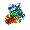
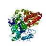
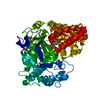
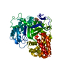
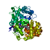
 Homo sapiens (human)
 X-RAY DIFFRACTION /
  Authors
 Citation
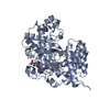
 Structure visualization
 Downloads & links
 Other downloads

 PDBj
PDBj
 Assembly
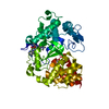
 unidentified baculovirus / Strain (production host): sf9


 Mass: 65.409 Da / Num. of mol.: 3 / Source method: obtained synthetically / Formula: Zn
 Mass: 24.305 Da / Num. of mol.: 2 / Source method: obtained synthetically / Formula: Mg
 Mass: 78.133 Da / Num. of mol.: 1 / Source method: obtained synthetically / Formula: C2H6OS / Comment: DMSO, precipitant*YM
 Mass: 452.508 Da / Num. of mol.: 1 / Source method: obtained synthetically / Formula: C19H32N8O5
 Sample preparation
 / Beamline: 21-ID-D / Wavelength: 1.0782 Å
 Processing