 Movie
Movie Controller

[English] 日本語
 Yorodumi
- PDB-5hm3: 2.25 Angstrom Resolution Crystal Structure of Long-chain-fatty-ac... -
+ Open data

- Basic information
| Entry | Database: PDB / ID: 5hm3 | ||||||
|---|---|---|---|---|---|---|---|
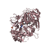
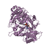
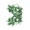
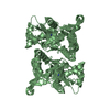
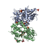
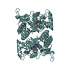
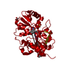
| Title | 2.25 Angstrom Resolution Crystal Structure of Long-chain-fatty-acid-AMP Ligase FadD32 from Mycobacterium tuberculosis in complex with Inhibitor 5'-O-[(11-phenoxyundecanoyl)sulfamoyl]adenosine | ||||||
 Components Components | Long-chain-fatty-acid--AMP ligase FadD32 | ||||||
 Keywords Keywords | LIGASE/LIGASE INHIBITOR / long-chain-fatty-acid--AMP ligase / FadD32 / Structural Genomics / Center for Structural Genomics of Infectious Diseases / CSGID / LIGASE-LIGASE INHIBITOR complex | ||||||
| Function / homology |  Function and homology information Function and homology informationlong-chain-fatty-acid-[acyl-carrier-protein] ligase / long-chain fatty acid [acyl-carrier-protein] ligase activity / adenylyltransferase activity / mycolate cell wall layer assembly / Actinobacterium-type cell wall biogenesis / lipid biosynthetic process / ligase activity / peptidoglycan-based cell wall / fatty acid biosynthetic process / ATP binding / plasma membrane Similarity search - Function | ||||||
| Biological species |   Mycobacterium tuberculosis (bacteria) Mycobacterium tuberculosis (bacteria) | ||||||
| Method |  X-RAY DIFFRACTION / SYNCHROTRON / SAD / Resolution: 2.25 Å X-RAY DIFFRACTION / SYNCHROTRON / SAD / Resolution: 2.25 Å | ||||||
 Authors Authors | Minasov, G. / Warwrzak, Z. / Kuhn, M.L. / Shuvalova, L. / Flores, K.J. / Wilson, D.J. / Grimes, K.D. / Aldrich, C.C. / Anderson, W.A. / Center for Structural Genomics of Infectious Diseases (CSGID) | ||||||
 Citation Citation | Journal: Acs Infect Dis. / Year: 2016 Title: Structure of the Essential Mtb FadD32 Enzyme: A Promising Drug Target for Treating Tuberculosis. Authors: Kuhn, M.L. / Alexander, E. / Minasov, G. / Page, H.J. / Warwrzak, Z. / Shuvalova, L. / Flores, K.J. / Wilson, D.J. / Shi, C. / Aldrich, C.C. / Anderson, W.F. | ||||||
| History |
|
- Structure visualization
| Structure viewer | Molecule: MolmilJmol/JSmol |
|---|
- Downloads & links
-Download
| PDBx/mmCIF format | 5hm3.cif.gz | 274.1 KB | Display | PDBx/mmCIF format |
|---|---|---|---|---|
| PDB format | pdb5hm3.ent.gz | 219.6 KB | Display | PDB format |
| PDBx/mmJSON format | 5hm3.json.gz | Tree view | PDBx/mmJSON format | |
| Others |  Other downloads Other downloads |
-Validation report
| Arichive directory | https://data.pdbj.org/pub/pdb/validation_reports/hm/5hm3ftp://data.pdbj.org/pub/pdb/validation_reports/hm/5hm3 | HTTPS FTP |
|---|
-Related structure data
| Similar structure data | |
|---|---|
| Other databases |


-Links
 PDBj
PDBj
- Assembly
| Deposited unit | 
| ||||||||
|---|---|---|---|---|---|---|---|---|---|
| 1 |
| ||||||||
| Unit cell |
|
-Components
-Protein , 1 types, 1 molecules A
| #1: Protein | Mass: 71887.539 Da / Num. of mol.: 1 Source method: isolated from a genetically manipulated source Source: (gene. exp.) Mycobacterium tuberculosis (strain ATCC 25618 / H37Rv) (bacteria)Strain: ATCC 25618 / H37Rv / Gene: fadD32, Rv3801c / Plasmid: pET28b / Production host: References: UniProt: O53580, Ligases; Forming carbon-sulfur bonds; Acid-thiol ligases |
|---|
-Non-polymers , 6 types, 140 molecules 


| #2: Chemical | ChemComp-649 /  Mass: 606.691 Da / Num. of mol.: 1 / Source method: obtained synthetically / Formula: C27H38N6O8S Mass: 606.691 Da / Num. of mol.: 1 / Source method: obtained synthetically / Formula: C27H38N6O8S | ||||||||
|---|---|---|---|---|---|---|---|---|---|
| #3: Chemical |  Mass: 35.453 Da / Num. of mol.: 2 / Source method: obtained synthetically / Formula: Cl Mass: 35.453 Da / Num. of mol.: 2 / Source method: obtained synthetically / Formula: Cl#4: Chemical |  Mass: 94.971 Da / Num. of mol.: 3 / Source method: obtained synthetically / Formula: PO4 Mass: 94.971 Da / Num. of mol.: 3 / Source method: obtained synthetically / Formula: PO4#5: Chemical | ChemComp-PEG / |  Mass: 106.120 Da / Num. of mol.: 1 / Source method: obtained synthetically / Formula: C4H10O3 Mass: 106.120 Da / Num. of mol.: 1 / Source method: obtained synthetically / Formula: C4H10O3#6: Chemical |  Mass: 92.094 Da / Num. of mol.: 2 / Source method: obtained synthetically / Formula: C3H8O3 Mass: 92.094 Da / Num. of mol.: 2 / Source method: obtained synthetically / Formula: C3H8O3#7: Water | ChemComp-HOH / | Mass: 18.015 Da / Num. of mol.: 131 / Source method: isolated from a natural source / Formula: H2O |
-Details
| Has protein modification | Y |
|---|
-Experimental details
-Experiment
| Experiment | Method: X-RAY DIFFRACTION / Number of used crystals: 1 |
|---|
- Sample preparation
| Crystal | Density Matthews: 2.02 Å3/Da / Density % sol: 39 % |
|---|---|
| Crystal grow | Temperature: 295 K / Method: vapor diffusion, sitting drop / pH: 7.6 Details: Protein: 6.0 mG/mL, 0.1M Pottasium chloride, 0.01 M Tris-HCL buffer pH 8.3, 5% glycerol, and 0.5 mM TCEP; Screen: Cubic Phase I (F10), 2.4 M Na/K phosphate pH 7.6 |
-Data collection
| Diffraction | Mean temperature: 100 K |
|---|---|
| Diffraction source | Source: SYNCHROTRON / Site: APS  / Beamline: 21-ID-D / Wavelength: 0.97875 Å / Beamline: 21-ID-D / Wavelength: 0.97875 Å |
| Detector | Type: MARMOSAIC 300 mm CCD / Detector: CCD / Date: Mar 20, 2014 / Details: Si(111) |
| Radiation | Monochromator: mirrors / Protocol: SINGLE WAVELENGTH / Monochromatic (M) / Laue (L): M / Scattering type: x-ray |
| Radiation wavelength | Wavelength: 0.97875 Å / Relative weight: 1 |
| Reflection | Resolution: 2.25→30 Å / Num. obs: 29261 / % possible obs: 99.9 % / Observed criterion σ(I): -3 / Redundancy: 4.9 % / Biso Wilson estimate: 49.5 Å2 / Rmerge(I) obs: 0.074 / Rsym value: 0.074 / Net I/σ(I): 18.4 |
| Reflection shell | Resolution: 2.25→2.29 Å / Redundancy: 4.9 % / Rmerge(I) obs: 0.583 / Mean I/σ(I) obs: 2.8 / % possible all: 100 |
- Processing
| Software |
| ||||||||||||||||||||||||||||||||||||||||||||||||||||||||||||||||||||||||||||||||||||||||||||||||||||||||||||||||||||||||||||||||||||||||||||||||||||||||||||||||||||||||||||||||||||||
|---|---|---|---|---|---|---|---|---|---|---|---|---|---|---|---|---|---|---|---|---|---|---|---|---|---|---|---|---|---|---|---|---|---|---|---|---|---|---|---|---|---|---|---|---|---|---|---|---|---|---|---|---|---|---|---|---|---|---|---|---|---|---|---|---|---|---|---|---|---|---|---|---|---|---|---|---|---|---|---|---|---|---|---|---|---|---|---|---|---|---|---|---|---|---|---|---|---|---|---|---|---|---|---|---|---|---|---|---|---|---|---|---|---|---|---|---|---|---|---|---|---|---|---|---|---|---|---|---|---|---|---|---|---|---|---|---|---|---|---|---|---|---|---|---|---|---|---|---|---|---|---|---|---|---|---|---|---|---|---|---|---|---|---|---|---|---|---|---|---|---|---|---|---|---|---|---|---|---|---|---|---|---|---|
| Refinement | Method to determine structure: SAD / Resolution: 2.25→29.36 Å / Cor.coef. Fo:Fc: 0.963 / Cor.coef. Fo:Fc free: 0.94 / SU B: 13.796 / SU ML: 0.173 / Cross valid method: THROUGHOUT / ESU R: 0.376 / ESU R Free: 0.238 / Details: HYDROGENS HAVE BEEN ADDED IN THE RIDING POSITIONS
| ||||||||||||||||||||||||||||||||||||||||||||||||||||||||||||||||||||||||||||||||||||||||||||||||||||||||||||||||||||||||||||||||||||||||||||||||||||||||||||||||||||||||||||||||||||||
| Solvent computation | Ion probe radii: 0.8 Å / Shrinkage radii: 0.8 Å / VDW probe radii: 1.2 Å | ||||||||||||||||||||||||||||||||||||||||||||||||||||||||||||||||||||||||||||||||||||||||||||||||||||||||||||||||||||||||||||||||||||||||||||||||||||||||||||||||||||||||||||||||||||||
| Displacement parameters | Biso mean: 62.991 Å2
| ||||||||||||||||||||||||||||||||||||||||||||||||||||||||||||||||||||||||||||||||||||||||||||||||||||||||||||||||||||||||||||||||||||||||||||||||||||||||||||||||||||||||||||||||||||||
| Refinement step | Cycle: 1 / Resolution: 2.25→29.36 Å
| ||||||||||||||||||||||||||||||||||||||||||||||||||||||||||||||||||||||||||||||||||||||||||||||||||||||||||||||||||||||||||||||||||||||||||||||||||||||||||||||||||||||||||||||||||||||
| Refine LS restraints |
|