Mass: 18.015 Da / Num. of mol.: 812 / Source method: isolated from a natural source / Formula: H2O
Has protein modification
Y
-
Experimental details
-
Experiment
Experiment
Method: X-RAY DIFFRACTION
-
Sample preparation
Crystal
Density Matthews: 2.98 Å3/Da / Density % sol: 58.69 %
Crystal grow
Temperature: 293 K / Method: vapor diffusion, sitting drop Details: 1/1 ratio of InlP 8.5 mg/ml in 10 mMTrisHCl pH 8.3, 1 mM TCEP and 0.2 M Calcium Chloride, 0.1 M TrisHCl pH 8, 20%(w/v) PEG 6000
Type: MARMOSAIC 300 mm CCD / Detector: CCD / Date: Jun 22, 2015
Radiation
Protocol: SINGLE WAVELENGTH / Monochromatic (M) / Laue (L): M / Scattering type: x-ray
Radiation wavelength
Wavelength: 0.97856 Å / Relative weight: 1
Reflection
Resolution: 1.4→30 Å / Num. obs: 180423 / % possible obs: 99.8 % / Redundancy: 5.6 % / Rmerge(I) obs: 0.079 / Rsym value: 0.079 / Net I/σ(I): 30.2
Reflection shell
Resolution: 1.4→1.42 Å / Redundancy: 5.3 % / Rmerge(I) obs: 0.653 / Mean I/σ(I) obs: 2.6 / % possible all: 100
-
Processing
Software
Name
Version
Classification
REFMAC
5.8.0107
refinement
HKL-3000
datareduction
HKL-3000
datascaling
HKL-3000
phasing
BLU-MAX
datacollection
Refinement
Method to determine structure: SAD / Resolution: 1.4→30 Å / Cor.coef. Fo:Fc: 0.97 / Cor.coef. Fo:Fc free: 0.965 / SU B: 1.501 / SU ML: 0.033 / Cross valid method: THROUGHOUT / ESU R: 0.051 / ESU R Free: 0.051 / Stereochemistry target values: MAXIMUM LIKELIHOOD / Details: HYDROGENS HAVE BEEN ADDED IN THE RIDING POSITIONS
Rfactor
Num. reflection
% reflection
Selection details
Rfree
0.176
4635
4.9 %
RANDOM
Rwork
0.15987
-
-
-
obs
0.16064
90409
99.75 %
-
Solvent computation
Ion probe radii: 0.8 Å / Shrinkage radii: 0.8 Å / VDW probe radii: 1.2 Å / Solvent model: MASK
 Movie
Movie Controller


 Open data
 Basic information
 Components
 Keywords
 Function and homology information
 Listeria monocytogenes EGD-e (bacteria)
 X-RAY DIFFRACTION /
  Authors
 Citation
 Structure visualization
 Downloads & links
 Other downloads
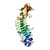


 PDBj
PDBj


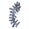
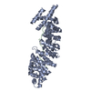
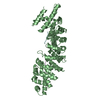
 Assembly

 Mass: 40.078 Da / Num. of mol.: 8 / Source method: obtained synthetically / Formula: Ca
 Mass: 35.453 Da / Num. of mol.: 1 / Source method: obtained synthetically / Formula: Cl
 Mass: 18.015 Da / Num. of mol.: 812 / Source method: isolated from a natural source / Formula: H2O
 Sample preparation
 / Beamline: 21-ID-G / Wavelength: 0.97856 Å
 Processing