 Movie
Movie Controller

[English] 日本語
 Yorodumi
- PDB-5hj1: Crystal structure of PDZ domain of pullulanase C protein of type ... -
+ Open data

- Basic information
| Entry | Database: PDB / ID: 5hj1 | ||||||
|---|---|---|---|---|---|---|---|
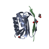
| Title | Crystal structure of PDZ domain of pullulanase C protein of type II secretion system from Klebsiella pneumoniae in complex with fatty acid | ||||||
 Components Components | Pullulanase C protein | ||||||
 Keywords Keywords | HYDROLASE / GspC protein / Structural Genomics / CSGID / Center for Structural Genomics of Infectious Diseases | ||||||
| Function / homology | PDZ domain / Pdz3 Domain / Roll / Mainly Beta / VACCENIC ACID / :  Function and homology information Function and homology information | ||||||
| Biological species |  Klebsiella pneumoniae subsp. pneumoniae NTUH-K2044 (bacteria) Klebsiella pneumoniae subsp. pneumoniae NTUH-K2044 (bacteria) | ||||||
| Method |  X-RAY DIFFRACTION / SYNCHROTRON / SAD / Resolution: 1.5 Å X-RAY DIFFRACTION / SYNCHROTRON / SAD / Resolution: 1.5 Å | ||||||
 Authors Authors | Filippova, E.V. / Minasov, G. / Shuvalova, L. / Kiryukhina, O. / Dubrovska, I. / Grimshaw, S. / Kwon, K. / Anderson, W.F. / Center for Structural Genomics of Infectious Diseases (CSGID) | ||||||
 Citation Citation | Journal: To Be Published Title: Crystal structure of PDZ domain of pullulanase C protein of type II secretion system from Klebsiella pneumoniae in complex with fatty acid Authors: Filippova, E.V. / Minasov, G. / Shuvalova, L. / Kiryukhina, O. / Dubrovska, I. / Grimshaw, S. / Kwon, K. / Anderson, W.F. / Center for Structural Genomics of Infectious Diseases (CSGID) | ||||||
| History |
|
- Structure visualization
| Structure viewer | Molecule: MolmilJmol/JSmol |
|---|
- Downloads & links
-Download
| PDBx/mmCIF format | 5hj1.cif.gz | 58.1 KB | Display | PDBx/mmCIF format |
|---|---|---|---|---|
| PDB format | pdb5hj1.ent.gz | 41.7 KB | Display | PDB format |
| PDBx/mmJSON format | 5hj1.json.gz | Tree view | PDBx/mmJSON format | |
| Others |  Other downloads Other downloads |
-Validation report
| Arichive directory | https://data.pdbj.org/pub/pdb/validation_reports/hj/5hj1ftp://data.pdbj.org/pub/pdb/validation_reports/hj/5hj1 | HTTPS FTP |
|---|
-Related structure data
| Similar structure data | |
|---|---|
| Other databases |


-Links
 PDBj
PDBj- Assembly
| Deposited unit | 
| ||||||||
|---|---|---|---|---|---|---|---|---|---|
| 1 |
| ||||||||
| Unit cell |
|
-Components
| #1: Protein | Mass: 11429.631 Da / Num. of mol.: 1 / Fragment: PDZ domain (UNP residues 181-280) Source method: isolated from a genetically manipulated source Source: (gene. exp.) Klebsiella pneumoniae subsp. pneumoniae NTUH-K2044 (bacteria)Gene: YP_002917874 / Plasmid: pMCSG53 / Production host: |
|---|---|
| #2: Chemical | ChemComp-VCA / 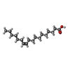  Mass: 282.461 Da / Num. of mol.: 1 / Source method: obtained synthetically / Formula: C18H34O2 Mass: 282.461 Da / Num. of mol.: 1 / Source method: obtained synthetically / Formula: C18H34O2 |
| #3: Water | ChemComp-HOH /  Mass: 18.015 Da / Num. of mol.: 78 / Source method: isolated from a natural source / Formula: H2O Mass: 18.015 Da / Num. of mol.: 78 / Source method: isolated from a natural source / Formula: H2O |
| Has protein modification | Y |
| Nonpolymer details | The ligand was identified based on the electron density maps and was not originally present in ...The ligand was identified based on the electron density maps and was not originally present in protein solution or crystallization conditions. Based on the results described by R. Mejia, et al., 1999 authors concluded that vaccenic acid is likely to bind to a protein. |
-Experimental details
-Experiment
| Experiment | Method: X-RAY DIFFRACTION / Number of used crystals: 1 |
|---|
- Sample preparation
| Crystal | Density Matthews: 2.32 Å3/Da / Density % sol: 47.04 % |
|---|---|
| Crystal grow | Temperature: 292 K / Method: vapor diffusion, sitting drop / Details: 4 M Formate |
-Data collection
| Diffraction | Mean temperature: 100 K |
|---|---|
| Diffraction source | Source: SYNCHROTRON / Site: APS  / Beamline: 21-ID-F / Wavelength: 0.97872 Å / Beamline: 21-ID-F / Wavelength: 0.97872 Å |
| Detector | Type: MARMOSAIC 225 mm CCD / Detector: CCD / Date: Dec 14, 2015 |
| Radiation | Monochromator: C(111) / Protocol: SINGLE WAVELENGTH / Monochromatic (M) / Laue (L): M / Scattering type: x-ray |
| Radiation wavelength | Wavelength: 0.97872 Å / Relative weight: 1 |
| Reflection | Resolution: 1.5→50 Å / Num. obs: 17852 / % possible obs: 99.6 % / Redundancy: 27.7 % / Rmerge(I) obs: 0.06 / Net I/σ(I): 87.8 |
| Reflection shell | Resolution: 1.5→1.53 Å / Redundancy: 28.3 % / Rmerge(I) obs: 0.57 / Mean I/σ(I) obs: 6.9 / % possible all: 100 |
- Processing
| Software |
| ||||||||||||||||||||||||||||||||||||||||||||||||||||||||||||||||||||||||||||||||||||||||||||||||||||||||||||||||||||||||||||||||||||||||||||||||||||||||||||||||||||||||||||||||||||||
|---|---|---|---|---|---|---|---|---|---|---|---|---|---|---|---|---|---|---|---|---|---|---|---|---|---|---|---|---|---|---|---|---|---|---|---|---|---|---|---|---|---|---|---|---|---|---|---|---|---|---|---|---|---|---|---|---|---|---|---|---|---|---|---|---|---|---|---|---|---|---|---|---|---|---|---|---|---|---|---|---|---|---|---|---|---|---|---|---|---|---|---|---|---|---|---|---|---|---|---|---|---|---|---|---|---|---|---|---|---|---|---|---|---|---|---|---|---|---|---|---|---|---|---|---|---|---|---|---|---|---|---|---|---|---|---|---|---|---|---|---|---|---|---|---|---|---|---|---|---|---|---|---|---|---|---|---|---|---|---|---|---|---|---|---|---|---|---|---|---|---|---|---|---|---|---|---|---|---|---|---|---|---|---|
| Refinement | Method to determine structure: SAD / Resolution: 1.5→42.42 Å / Cor.coef. Fo:Fc: 0.959 / Cor.coef. Fo:Fc free: 0.95 / SU B: 2.181 / SU ML: 0.041 / Cross valid method: THROUGHOUT / ESU R: 0.077 / ESU R Free: 0.079 / Stereochemistry target values: MAXIMUM LIKELIHOOD Details: HYDROGENS HAVE BEEN ADDED IN THE RIDING POSITIONS. The ligand was identified based on the electron density maps and was not originally present in protein solution or crystallization ...Details: HYDROGENS HAVE BEEN ADDED IN THE RIDING POSITIONS. The ligand was identified based on the electron density maps and was not originally present in protein solution or crystallization conditions. Based on the results described by R. Mejia, et al., 1999 authors concluded that vaccenic acid is likely to bind to a protein.
| ||||||||||||||||||||||||||||||||||||||||||||||||||||||||||||||||||||||||||||||||||||||||||||||||||||||||||||||||||||||||||||||||||||||||||||||||||||||||||||||||||||||||||||||||||||||
| Solvent computation | Ion probe radii: 0.8 Å / Shrinkage radii: 0.8 Å / VDW probe radii: 1.2 Å / Solvent model: MASK | ||||||||||||||||||||||||||||||||||||||||||||||||||||||||||||||||||||||||||||||||||||||||||||||||||||||||||||||||||||||||||||||||||||||||||||||||||||||||||||||||||||||||||||||||||||||
| Displacement parameters | Biso mean: 32.17 Å2
| ||||||||||||||||||||||||||||||||||||||||||||||||||||||||||||||||||||||||||||||||||||||||||||||||||||||||||||||||||||||||||||||||||||||||||||||||||||||||||||||||||||||||||||||||||||||
| Refinement step | Cycle: LAST / Resolution: 1.5→42.42 Å
| ||||||||||||||||||||||||||||||||||||||||||||||||||||||||||||||||||||||||||||||||||||||||||||||||||||||||||||||||||||||||||||||||||||||||||||||||||||||||||||||||||||||||||||||||||||||
| Refine LS restraints |
|