 Movie
Movie Controller


+ Open data

- Basic information
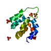
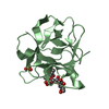
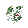
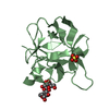
| Entry | Database: PDB / ID: 1odd | ||||||
|---|---|---|---|---|---|---|---|
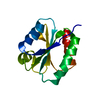
| Title | OMPR C-TERMINAL DOMAIN (OMPR-C) FROM ESCHERICHIA COLI | ||||||
 Components Components | TRANSCRIPTIONAL REGULATORY PROTEIN OMPR | ||||||
 Keywords Keywords | GENE REGULATORY PROTEIN / POSITIVE RESPONSE REGULATOR / DNA BINDING DOMAIN | ||||||
| Function / homology |  Function and homology information Function and homology informationphosphorelay response regulator activity / phosphorelay signal transduction system / protein-DNA complex / transcription cis-regulatory region binding / regulation of DNA-templated transcription / positive regulation of DNA-templated transcription / cytosol Similarity search - Function | ||||||
| Biological species |  | ||||||
| Method |  X-RAY DIFFRACTION / SYNCHROTRON / MAD WITH SE-MET MUTANT / Resolution: 2.2 Å X-RAY DIFFRACTION / SYNCHROTRON / MAD WITH SE-MET MUTANT / Resolution: 2.2 Å | ||||||
 Authors Authors | Kondou, H. / Nakagawa, A. / Tanaka, I. | ||||||
 Citation Citation | Journal: Nat.Struct.Biol. / Year: 1997 Title: Escherichia coli positive regulator OmpR has a large loop structure at the putative RNA polymerase interaction site. Authors: Kondo, H. / Nakagawa, A. / Nishihira, J. / Nishimura, Y. / Mizuno, T. / Tanaka, I. #1: Journal: J.Mol.Biol. / Year: 1994Title: Crystallization and X-Ray Studies of the DNA-Binding Domain of OmpR Protein, a Positive Regulator Involved in Activation of Osmoregulatory Genes in Escherichia Coli Authors: Kondo, H. / Miyaji, T. / Suzuki, M. / Tate, S. / Mizuno, T. / Nishimura, Y. / Tanaka, I. | ||||||
| History |
|
- Structure visualization
| Structure viewer | Molecule: MolmilJmol/JSmol |
|---|
- Downloads & links
-Download
| PDBx/mmCIF format | 1odd.cif.gz | 32.6 KB | Display | PDBx/mmCIF format |
|---|---|---|---|---|
| PDB format | pdb1odd.ent.gz | 21.9 KB | Display | PDB format |
| PDBx/mmJSON format | 1odd.json.gz | Tree view | PDBx/mmJSON format | |
| Others |  Other downloads Other downloads |
-Validation report
| Arichive directory | https://data.pdbj.org/pub/pdb/validation_reports/od/1oddftp://data.pdbj.org/pub/pdb/validation_reports/od/1odd | HTTPS FTP |
|---|
-Related structure data
| Similar structure data |
|---|


-Links
 PDBj
PDBj

- Assembly
| Deposited unit | 
| ||||||||
|---|---|---|---|---|---|---|---|---|---|
| 1 |
| ||||||||
| Unit cell |
|
-Components
| #1: Protein | Mass: 13405.311 Da / Num. of mol.: 1 / Fragment: DNA-BINDING DOMAIN Source method: isolated from a genetically manipulated source Source: (gene. exp.) |
|---|---|
| #2: Water | ChemComp-HOH /  Mass: 18.015 Da / Num. of mol.: 52 / Source method: isolated from a natural source / Formula: H2O Mass: 18.015 Da / Num. of mol.: 52 / Source method: isolated from a natural source / Formula: H2O |
-Experimental details
-Experiment
| Experiment | Method: X-RAY DIFFRACTION / Number of used crystals: 1 |
|---|
- Sample preparation
| Crystal | Density Matthews: 2.4 Å3/Da / Density % sol: 47 % Description: DATA WERE COLLECTED USING THE WEISSENBERG METHOD. | |||||||||||||||
|---|---|---|---|---|---|---|---|---|---|---|---|---|---|---|---|---|
| Crystal grow | pH: 6 Details: PROTEIN WAS CRYSTALLIZED FROM 0.1M SODIUM CACODYLATE, 0.2M SODIUM ACETATE, 20%(W/V) PEG8000, PH 6.0; THEN SOAKED IN 35% PEG8000 WITH CRYSTALLIZATION BUFFER. | |||||||||||||||
| Crystal grow | *PLUS Temperature: 18 ℃ / Method: vapor diffusion, hanging drop / Details: Kondo, H., (1994) J.Mol.Biol., 235, 780. | |||||||||||||||
| Components of the solutions | *PLUS
|
-Data collection
| Diffraction | Mean temperature: 100 K |
|---|---|
| Diffraction source | Source: SYNCHROTRON / Site: Photon Factory  / Beamline: BL-18B / Wavelength: 1 / Beamline: BL-18B / Wavelength: 1 |
| Detector | Type: FUJI / Detector: IMAGE PLATE / Date: May 1, 1996 / Details: MIRROR, COLLIMATOR |
| Radiation | Monochromator: SI(111) / Monochromatic (M) / Laue (L): M / Scattering type: x-ray |
| Radiation wavelength | Wavelength: 1 Å / Relative weight: 1 |
| Reflection | Resolution: 2.2→20 Å / Num. obs: 6153 / % possible obs: 98.1 % / Observed criterion σ(I): 4 / Redundancy: 5.1 % / Rmerge(I) obs: 0.072 / Net I/σ(I): 8 |
| Reflection shell | Resolution: 2.2→2.32 Å / Redundancy: 5.2 % / Rmerge(I) obs: 0.307 / Mean I/σ(I) obs: 1.9 / % possible all: 96.3 |
| Reflection | *PLUS Num. measured all: 31412 |
| Reflection shell | *PLUS % possible obs: 96.3 % |
- Processing
| Software |
| |||||||||||||||||||||
|---|---|---|---|---|---|---|---|---|---|---|---|---|---|---|---|---|---|---|---|---|---|---|
| Refinement | Method to determine structure: MAD WITH SE-MET MUTANT / Resolution: 2.2→10 Å / Cross valid method: THROUGHOUT / σ(F): 0 / Details: PROGRAM X-PLOR BY BRUNGER ALSO WAS USED.
| |||||||||||||||||||||
| Refinement step | Cycle: LAST / Resolution: 2.2→10 Å
| |||||||||||||||||||||
| Software | *PLUS Name: REFMAC / Classification: refinement | |||||||||||||||||||||
| Refine LS restraints | *PLUS
|
