 Movie
Movie Controller

[English] 日本語
 Yorodumi
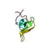
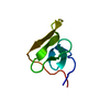
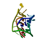
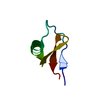
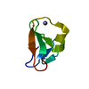
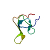
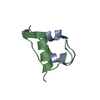
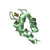
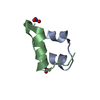
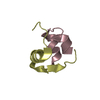
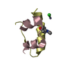
- PDB-5hir: SOLUTION STRUCTURE OF RECOMBINANT HIRUDIN AND THE LYS-47 (RIGHT A... -
+ Open data

- Basic information
| Entry | Database: PDB / ID: 5hir | ||||||
|---|---|---|---|---|---|---|---|
| Title | SOLUTION STRUCTURE OF RECOMBINANT HIRUDIN AND THE LYS-47 (RIGHT ARROW) GLU MUTANT. A NUCLEAR MAGNETIC RESONANCE AND HYBRID DISTANCE GEOMETRY-DYNAMICAL SIMULATED ANNEALING STUDY | ||||||
 Components Components | HIRUDIN VARIANT-1 | ||||||
 Keywords Keywords | COAGULATION INHIBITOR | ||||||
| Function / homology |  Function and homology information Function and homology informationnegative regulation of serine-type peptidase activity / serine-type endopeptidase inhibitor activity / toxin activity / extracellular space Similarity search - Function | ||||||
| Biological species |  Hirudo medicinalis (medicinal leech) Hirudo medicinalis (medicinal leech) | ||||||
| Method | SOLUTION NMR | ||||||
 Authors Authors | Clore, G.M. / Gronenborn, A.M. | ||||||
 Citation Citation | Journal: Biochemistry / Year: 1989 Title: Solution structure of recombinant hirudin and the Lys-47----Glu mutant: a nuclear magnetic resonance and hybrid distance geometry-dynamical simulated annealing study. Authors: Folkers, P.J. / Clore, G.M. / Driscoll, P.C. / Dodt, J. / Kohler, S. / Gronenborn, A.M. | ||||||
| History |
|
- Structure visualization
| Structure viewer | Molecule: MolmilJmol/JSmol |
|---|
- Downloads & links
-Download
| PDBx/mmCIF format | 5hir.cif.gz | 25.5 KB | Display | PDBx/mmCIF format |
|---|---|---|---|---|
| PDB format | pdb5hir.ent.gz | 16.6 KB | Display | PDB format |
| PDBx/mmJSON format | 5hir.json.gz | Tree view | PDBx/mmJSON format | |
| Others |  Other downloads Other downloads |
-Validation report
| Arichive directory | https://data.pdbj.org/pub/pdb/validation_reports/hi/5hirftp://data.pdbj.org/pub/pdb/validation_reports/hi/5hir | HTTPS FTP |
|---|
-Related structure data


-Links
 PDBj
PDBj
- Assembly
| Deposited unit | 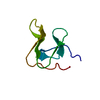
| |||||||||
|---|---|---|---|---|---|---|---|---|---|---|
| 1 |
| |||||||||
| NMR ensembles |
|
-Components
| #1: Protein | Mass: 6973.505 Da / Num. of mol.: 1 Source method: isolated from a genetically manipulated source Source: (gene. exp.) Hirudo medicinalis (medicinal leech) / References: UniProt: P01050 |
|---|---|
| Has protein modification | Y |
-Experimental details
-Experiment
| Experiment | Method: SOLUTION NMR |
|---|
- Sample preparation
| Crystal grow | *PLUS Method: other / Details: NMR |
|---|
- Processing
| Software |
| ||||||||
|---|---|---|---|---|---|---|---|---|---|
| NMR software | Name:  X-PLOR / Developer: BRUNGER / Classification: refinement X-PLOR / Developer: BRUNGER / Classification: refinement | ||||||||
| Refinement | Software ordinal: 1 Details: THE METHOD USED TO DETERMINE AND REFINE THE STRUCTURE IS THE HYBRID METRIC MATRIX DISTANCE GEOMETRY-DYNAMICAL SIMULATED ANNEALING METHOD (M.NILGES, G.M.CLORE, A.M. GRONENBORN, FEBS LETT. ...Details: THE METHOD USED TO DETERMINE AND REFINE THE STRUCTURE IS THE HYBRID METRIC MATRIX DISTANCE GEOMETRY-DYNAMICAL SIMULATED ANNEALING METHOD (M.NILGES, G.M.CLORE, A.M. GRONENBORN, FEBS LETT. 229, 317-324 (1988)) USING THE PROGRAM XPLOR (A.T. BRUENGER, YALE UNIVERSITY, CT 06511). STRUCTURAL STATISTICS RMS DEVIATION FROM EXPERIMENTAL RESTRAINTS *(1)* RESTRAINT TYPE NUMBER OF RESTRAINTS RMS (ANGSTROMS) ALL 701 0.075 INTERRESIDUE SHORT RANGE 242 0.094 INTERRESIDUE LONG RANGE 208 0.057 INTRARESIDUE 235 0.067 HBOND *(2)* 16 0.052 POTENTIAL ENERGY TERMS TYPE ENERGY (KCAL/MOL) F(NOE) *(3)* 196 F(TOR) *(4)* 14 F(REPEL) *(5)* 45 LENNARD-JONES VAN DER WAALS ENERGY (E(L-J)) CALCULATED USING THE *CHARMM* EMPIRICAL ENERGY FUNCTION IS -73 KCAL/MOL. DEVIATIONS FROM IDEALIZED GEOMETRY *(6)* TYPE TOTAL NUMBER RMS DEVIATION BONDS 676 0.012 (ANGSTROMS) ANGLES 1223 1.814 (DEGREES) IMPROPERS 131 0.597 (DEGREES) NOTES. *(1)* THE RMS DEVIATION FROM THE EXPERIMENTAL RESTRAINTS ARE CALCULATED WITH RESPECT TO THE UPPER AND LOWER LIMITS OF THE DISTANCE RESTRAINTS. NONE OF THE STRUCTURES EXHIBITED VIOLATIONS GREATER THAN 0.5 ANGSTROMS. *(2)* FOR EACH BACKBONE HYDROGEN BOND THERE ARE TWO RESTRAINTS - R(NH-O) .LT. 2.3 ANGSTROMS AND R(N-O) .LT. 3.3 ANGSTROMS. THE LOWER LIMITS ARE GIVEN BY THE SUM OF THE VAN DER WAALS RADII OF THE RELEVANT ATOMS. *(3)* THE VALUES OF THE SQUARE-WELL NOE POTENTIAL F(NOE) ARE CALCULATED WITH A FORCE CONSTANT OF 50 KCAL/MOL/ANGSTROM**2. *(4)* THE VALUES OF F(PHI) ARE CALCULATED WITH A FORCE CONSTANT OF 200 KCAL/MOL/RAD**2. F(PHI) IS A SQUARE-WELL DIHEDRAL POTENTIAL WHICH IS USED TO RESTRICT THE RANGES OF 33 PHI ,24 PSI AND 25 CHI1 TORSION ANGLES. *(5)* THE VALUE OF THE VAN DER WAALS REPULSION TERM F(REPEL) IS CALCULATED WITH A FORCE CONSTANT OF 4 KCAL/MOL/ANGSTROM**4 WITH THE HARD SPHERE VAN DER WAALS RADII SET TO 0.8 TIMES THE STANDARD VALUES USED IN THE *CHARMM* EMPIRICAL ENERGY FUNCTION. *(6)* THE IMPROPER TERMS SERVE TO MAINTAIN PLANARITY AND APPROPRIATE CHIRALITY. THEY ALSO MAINTAIN THE PEPTIDE BONDS OF ALL RESIDUES (WITH THE EXCEPTION OF PROLINES) IN THE TRANS CONFORMATION. IN THE DYNAMICAL SIMULATED ANNEALING CALCULATIONS, THE RESTRAINTS FOR THE DISULFIDE BRIDGES ARE INCLUDED IN THE BOND AND ANGLE TERMS. A TOTAL OF 32 STRUCTURES CONSISTENT WITH THE NMR DATA WERE CALCULATED. THIS ENTRY REPRESENTS THE COORDINATES OBTAINED BY AVERAGING THE COORDINATES OF THE INDIVIDUAL STRUCTURES AND SUBJECTING THE RESULTING COORDINATES TO FURTHER RESTRAINED MINIMIZATION. THE COORDINATES OF THE 32 STRUCTURES ARE GIVEN IN THE PROTEIN DATA BANK ENTRY *2HIR*. THE THERMAL PARAMETERS GIVEN IN THIS ENTRY REPRESENT THE ATOMIC RMS DEVIATION OF THE INDIVIDUAL STRUCTURES ABOUT THE MEAN COORDINATE POSITIONS. ONLY RESIDUES 1-49 ARE WELL DEFINED. RESIDUES 50-65 FORM A DISORDERED C-TERMINAL TAIL. | ||||||||
| NMR ensemble | Conformers submitted total number: 1 |
