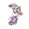
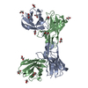
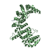
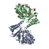
Entry Database : PDB / ID : 5hbzTitle Structure of EAV NSP11 K170A mutant at 3.10A Non-structural protein 11 Keywords / / / / / / Function / homology Function Domain/homology Component
/ / / / / / / / / / / / / / / / / / / / / / / / / / / / / / / / / / / / / / / / / / / / / / / / / / / / / / / / / / / / / / / / / / / / / / / / / / / / / / / / / / / / / / / / / / / / / / / / / / Biological species Method / / / Resolution : 3.1 Å Authors Zhang, M.F. / Chen, Z.Z. Journal : J. Virol. / Year : 2017Title : Structural Biology of the Arterivirus nsp11 Endoribonucleases.Authors : Zhang, M. / Li, X. / Deng, Z. / Chen, Z. / Liu, Y. / Gao, Y. / Wu, W. / Chen, Z. History Deposition Jan 4, 2016 Deposition site / Processing site Revision 1.0 Oct 12, 2016 Provider / Type Revision 1.1 Nov 16, 2016 Group Revision 1.2 Jan 18, 2017 Group Revision 1.3 Nov 8, 2023 Group / Database references / Refinement descriptionCategory chem_comp_atom / chem_comp_bond ... chem_comp_atom / chem_comp_bond / database_2 / pdbx_initial_refinement_model Item / _database_2.pdbx_database_accession
Show all Show less
 Movie
Movie Controller


 Open data
 Basic information
 Components
 Keywords
 Function and homology information
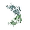
 Equine arteritis virus Bucyrus
 X-RAY DIFFRACTION /
  Authors
 Citation
 Structure visualization
 Downloads & links
 Other downloads


 PDBj
PDBj Assembly
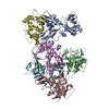


 Sample preparation
 / Beamline: BL17U / Wavelength: 1 Å
 Processing