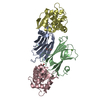
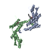
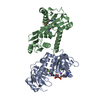
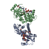
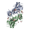
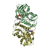
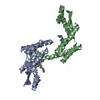
[English] 日本語
Yorodumi
- PDB-5eyi: Structure of PRRSV apo-NSP11 at 2.16A -

+
Open data


ID or keywords:

Loading...

-
Basic information

Entry
Database: PDB / ID: 5eyi
TitleStructure of PRRSV apo-NSP11 at 2.16A
ComponentsNon-structural protein 11
KeywordsHYDROLASE / Non-structural protein 11 / Nsp11 / Beta Interferon Antagonist / Endoribonuclease
Function / homology
Function and homology information


: / Hydrolases; Acting on peptide bonds (peptidases); Serine endopeptidases / host cell membrane / symbiont-mediated suppression of host JAK-STAT cascade via inhibition of STAT1 activity / Lyases; Phosphorus-oxygen lyases / symbiont-mediated suppression of host ISG15-protein conjugation / symbiont-mediated perturbation of host ubiquitin-like protein modification / endonuclease activity / DNA helicase / ubiquitinyl hydrolase 1 ...: / Hydrolases; Acting on peptide bonds (peptidases); Serine endopeptidases / host cell membrane / symbiont-mediated suppression of host JAK-STAT cascade via inhibition of STAT1 activity / Lyases; Phosphorus-oxygen lyases / symbiont-mediated suppression of host ISG15-protein conjugation / symbiont-mediated perturbation of host ubiquitin-like protein modification / endonuclease activity / DNA helicase / ubiquitinyl hydrolase 1 / cysteine-type deubiquitinase activity / Hydrolases; Acting on peptide bonds (peptidases); Cysteine endopeptidases / RNA helicase activity / host cell perinuclear region of cytoplasm / viral protein processing / lyase activity / symbiont-mediated suppression of host type I interferon-mediated signaling pathway / RNA helicase / RNA-directed RNA polymerase / viral RNA genome replication / cysteine-type endopeptidase activity / serine-type endopeptidase activity / RNA-dependent RNA polymerase activity / DNA-templated transcription / host cell nucleus / ATP hydrolysis activity / proteolysis / RNA binding / zinc ion binding / ATP binding / membrane
Similarity search - Function
Nonstructural protein 10, 1B domain, arterivirus / Replicase polyprotein 1ab, peptidase C33-associated domain / Peptidase_C33-associated domain / Arterivirus polyprotein, nsp2, immunogenic region / Arterivirus papain-like cysteine protease beta (PCPbeta) domain superfamily / Arterivirus papain-like cysteine protease alpha (PCPalpha) domain superfamily / Porcine arterivirus-type cysteine proteinase alpha / Equine arteritis virus putative proteinase / Immunogenic region of nsp2 protein of arterivirus polyprotein / Nonstructural protein 10, zinc-binding domain, arterivirus ...Nonstructural protein 10, 1B domain, arterivirus / Replicase polyprotein 1ab, peptidase C33-associated domain / Peptidase_C33-associated domain / Arterivirus polyprotein, nsp2, immunogenic region / Arterivirus papain-like cysteine protease beta (PCPbeta) domain superfamily / Arterivirus papain-like cysteine protease alpha (PCPalpha) domain superfamily / Porcine arterivirus-type cysteine proteinase alpha / Equine arteritis virus putative proteinase / Immunogenic region of nsp2 protein of arterivirus polyprotein / Nonstructural protein 10, zinc-binding domain, arterivirus / NSP11, NendoU domain, arterivirus / NSP11, N-terminal, arterivirus / Arteriviridae zinc-binding (AV ZBD) domain profile. / Arterivirus papain-like cysteine protease alpha (PCPalpha) domain / Arterivirus Nsp2, peptidase C33 / Equine arteritis virus peptidase S32 / Serine protease, chymotrypsin-like serine protease, C-terminal / Arterivirus NSP4 peptidase domain / Arterivirus papain-like cysteine protease beta (PCPbeta) domain / Arterivirus nonstructural protein 7 alpha / Arterivirus nsp7 alpha superfamily / Equine arterivirus Nsp2-type cysteine proteinase / Equine arteritis virus serine endopeptidase S32 / Arterivirus nonstructural protein 7 alpha / Arterivirus nsp4 proteinase domain profile. / Arterivirus nsp2 cysteine protease (AV CP) domain profile. / Arterivirus papain-like cysteine protease alpha (PCPalpha) domain profile. / Arterivirus papain-like cysteine protease beta (PCPbeta) domain profile. / Viral (Superfamily 1) RNA helicase / Arterivirus Nsp11 N-terminal/Coronavirus NSP15 middle domain / Arterivirus Nsp11 N-terminal/coronavirus NSP15 middle (AV-Nsp11N/CoV-Nsp15M) domain profile. / Nidoviral uridylate-specific endoribonuclease (NendoU) domain profile. / Nidovirus RdRp-associated nucleotidyl transferase (NiRAN) domain / Nidovirus RdRp-associated nucleotidyl transferase (NiRAN) domain profile. / Endoribonuclease EndoU-like / NendoU domain, nidovirus / Coronavirus replicase NSP15, uridylate-specific endoribonuclease / (+) RNA virus helicase core domain / (+)RNA virus helicase core domain profile. / RNA-directed RNA polymerase, C-terminal domain / Viral RNA-dependent RNA polymerase / RNA-directed RNA polymerase, catalytic domain / RdRp of positive ssRNA viruses catalytic domain profile. / Peptidase S1, PA clan, chymotrypsin-like fold / Peptidase S1, PA clan / DNA/RNA polymerase superfamily / P-loop containing nucleoside triphosphate hydrolase
Similarity search - Domain/homology
Replicase polyprotein 1ab
Similarity search - Component
Biological speciesPRRSV 16244B (virus)
MethodX-RAY DIFFRACTION / SYNCHROTRON / SAD / Resolution: 2.16 Å
AuthorsZhang, M.F. / Chen, Z.
CitationJournal: J. Virol. / Year: 2017
Title: Structural Biology of the Arterivirus nsp11 Endoribonucleases.
Authors: Zhang, M. / Li, X. / Deng, Z. / Chen, Z. / Liu, Y. / Gao, Y. / Wu, W. / Chen, Z.
History
DepositionNov 25, 2015Deposition site: RCSB / Processing site: PDBJ
Revision 1.0Oct 12, 2016Provider: repository / Type: Initial release
Revision 1.1Nov 16, 2016Group: Database references
Revision 1.2Jan 18, 2017Group: Database references
Revision 1.3Mar 20, 2024Group: Data collection / Database references / Derived calculations
Category: chem_comp_atom / chem_comp_bond ...chem_comp_atom / chem_comp_bond / database_2 / pdbx_struct_special_symmetry
Item: _database_2.pdbx_DOI / _database_2.pdbx_database_accession

-
Structure visualization

Structure viewerMolecule:
MolmilJmol/JSmol

Downloads & links

-
Assembly

Deposited unit
A: Non-structural protein 11
B: Non-structural protein 11
hetero molecules


Theoretical massNumber of molelcules
Total (without water)50,1775
Polymers49,9252
Non-polymers2523
Water7,927440
1


  • Idetical with deposited unit
  • defined by author
TypeNameSymmetry operationNumber
identity operation1_555x,y,z1
Buried area1990 Å2
ΔGint-14 kcal/mol
Surface area20730 Å2
Unit cell
Length a, b, c (Å)75.299, 75.299, 199.489
Angle α, β, γ (deg.)90.00, 90.00, 90.00
Int Tables number92
Space group name H-MP41212
Components on special symmetry positions
IDModelComponents
11B-346-

HOH

-
Components

#1: Protein Non-structural protein 11


Mass: 24962.703 Da / Num. of mol.: 2 / Mutation: K173A
Source method: isolated from a genetically manipulated source
Source: (gene. exp.) PRRSV 16244B (virus) / Strain: 16244B / Gene: rep, 1a-1b / Production host: Escherichia coli (E. coli) / References: UniProt: Q9YN02
#2: Chemical ChemComp-SO4 / SULFATE ION


Mass: 96.063 Da / Num. of mol.: 1 / Source method: obtained synthetically / Formula: SO4
#3: Chemical ChemComp-CL / CHLORIDE ION


Mass: 35.453 Da / Num. of mol.: 1 / Source method: obtained synthetically / Formula: Cl
#4: Chemical ChemComp-PG0 / 2-(2-METHOXYETHOXY)ETHANOL / PEG 6000


Mass: 120.147 Da / Num. of mol.: 1 / Source method: obtained synthetically / Formula: C5H12O3 / Comment: inhibitor, precipitant*YM
#5: Water ChemComp-HOH / water


Mass: 18.015 Da / Num. of mol.: 440 / Source method: isolated from a natural source / Formula: H2O

-
Experimental details

-
Experiment

ExperimentMethod: X-RAY DIFFRACTION

-
Sample preparation

CrystalDensity Matthews: 2.83 Å3/Da / Density % sol: 56.57 %
Crystal growTemperature: 277 K / Method: vapor diffusion, hanging drop / pH: 8 / Details: PEG

-
Data collection

DiffractionMean temperature: 100 K
Diffraction sourceSource: SYNCHROTRON / Site: SSRF / Beamline: BL18U1 / Wavelength: 0.9793 Å
DetectorType: ADSC QUANTUM 315 / Detector: CCD / Date: Sep 17, 2015
RadiationProtocol: SINGLE WAVELENGTH / Monochromatic (M) / Laue (L): M / Scattering type: x-ray
Radiation wavelengthWavelength: 0.9793 Å / Relative weight: 1
ReflectionResolution: 2.16→50 Å / Num. all: 31952 / Num. obs: 31822 / % possible obs: 99.6 % / Redundancy: 7.7 % / Net I/σ(I): 19
Reflection shellResolution: 2.16→2.2 Å / Redundancy: 7.9 % / Mean I/σ(I) obs: 1.5 / % possible all: 99.9

-
Processing

Software
NameVersionClassification
REFMAC5.8.0103refinement
HKL-2000data reduction
HKL-2000data scaling
HKL-2000phasing
RefinementMethod to determine structure: SAD / Resolution: 2.16→50 Å / Cor.coef. Fo:Fc: 0.953 / Cor.coef. Fo:Fc free: 0.937 / SU B: 5.102 / SU ML: 0.125 / Cross valid method: THROUGHOUT / ESU R: 0.21 / ESU R Free: 0.167 / Stereochemistry target values: MAXIMUM LIKELIHOOD / Details: HYDROGENS HAVE BEEN ADDED IN THE RIDING POSITIONS
RfactorNum. reflection% reflectionSelection details
Rfree0.21028 1607 5.1 %RANDOM
Rwork0.18441 ---
obs0.18578 29712 98.19 %-
Solvent computationIon probe radii: 0.8 Å / Shrinkage radii: 0.8 Å / VDW probe radii: 1.2 Å / Solvent model: MASK
Displacement parametersBiso mean: 29.309 Å2
Baniso -1Baniso -2Baniso -3
1-0.02 Å20 Å20 Å2
2--0.02 Å2-0 Å2
3----0.04 Å2
Refinement stepCycle: 1 / Resolution: 2.16→50 Å
ProteinNucleic acidLigandSolventTotal
Num. atoms3369 0 14 440 3823
Refine LS restraints
Refine-IDTypeDev idealDev ideal targetNumber
X-RAY DIFFRACTIONr_bond_refined_d0.0070.0193477
X-RAY DIFFRACTIONr_bond_other_d0.0020.023285
X-RAY DIFFRACTIONr_angle_refined_deg1.3371.9684735
X-RAY DIFFRACTIONr_angle_other_deg0.90137578
X-RAY DIFFRACTIONr_dihedral_angle_1_deg6.7095429
X-RAY DIFFRACTIONr_dihedral_angle_2_deg28.83622.632133
X-RAY DIFFRACTIONr_dihedral_angle_3_deg14.43515545
X-RAY DIFFRACTIONr_dihedral_angle_4_deg13.4271519
X-RAY DIFFRACTIONr_chiral_restr0.0770.2532
X-RAY DIFFRACTIONr_gen_planes_refined0.0050.0213822
X-RAY DIFFRACTIONr_gen_planes_other0.0010.02774
X-RAY DIFFRACTIONr_nbd_refined
X-RAY DIFFRACTIONr_nbd_other
X-RAY DIFFRACTIONr_nbtor_refined
X-RAY DIFFRACTIONr_nbtor_other
X-RAY DIFFRACTIONr_xyhbond_nbd_refined
X-RAY DIFFRACTIONr_xyhbond_nbd_other
X-RAY DIFFRACTIONr_metal_ion_refined
X-RAY DIFFRACTIONr_metal_ion_other
X-RAY DIFFRACTIONr_symmetry_vdw_refined
X-RAY DIFFRACTIONr_symmetry_vdw_other
X-RAY DIFFRACTIONr_symmetry_hbond_refined
X-RAY DIFFRACTIONr_symmetry_hbond_other
X-RAY DIFFRACTIONr_symmetry_metal_ion_refined
X-RAY DIFFRACTIONr_symmetry_metal_ion_other
X-RAY DIFFRACTIONr_mcbond_it1.2452.791728
X-RAY DIFFRACTIONr_mcbond_other1.2452.7891727
X-RAY DIFFRACTIONr_mcangle_it2.1164.1732153
X-RAY DIFFRACTIONr_mcangle_other2.1154.1742154
X-RAY DIFFRACTIONr_scbond_it1.5382.9981749
X-RAY DIFFRACTIONr_scbond_other1.5372.9991750
X-RAY DIFFRACTIONr_scangle_it
X-RAY DIFFRACTIONr_scangle_other2.64.3992582
X-RAY DIFFRACTIONr_long_range_B_refined5.33523.5064046
X-RAY DIFFRACTIONr_long_range_B_other5.16423.2243969
X-RAY DIFFRACTIONr_rigid_bond_restr
X-RAY DIFFRACTIONr_sphericity_free
X-RAY DIFFRACTIONr_sphericity_bonded
LS refinement shellResolution: 2.156→2.212 Å / Total num. of bins used: 20
RfactorNum. reflection% reflection
Rfree0.311 99 -
Rwork0.262 1863 -
obs--84.97 %

+
About Yorodumi

-
News

-
Feb 9, 2022. New format data for meta-information of EMDB entries

New format data for meta-information of EMDB entries

  • Version 3 of the EMDB header file is now the official format.
  • The previous official version 1.9 will be removed from the archive.

Related info.:EMDB header

External links:wwPDB to switch to version 3 of the EMDB data model

-
Aug 12, 2020. Covid-19 info

Covid-19 info

URL: https://pdbjlvh1.pdbj.org/emnavi/covid19.php

New page: Covid-19 featured information page in EM Navigator.

Related info.:Covid-19 info / Mar 5, 2020. Novel coronavirus structure data

+
Mar 5, 2020. Novel coronavirus structure data

Novel coronavirus structure data

Related info.:Yorodumi Speices / Aug 12, 2020. Covid-19 info

External links:COVID-19 featured content - PDBj / Molecule of the Month (242):Coronavirus Proteases

+
Jan 31, 2019. EMDB accession codes are about to change! (news from PDBe EMDB page)

EMDB accession codes are about to change! (news from PDBe EMDB page)

  • The allocation of 4 digits for EMDB accession codes will soon come to an end. Whilst these codes will remain in use, new EMDB accession codes will include an additional digit and will expand incrementally as the available range of codes is exhausted. The current 4-digit format prefixed with “EMD-” (i.e. EMD-XXXX) will advance to a 5-digit format (i.e. EMD-XXXXX), and so on. It is currently estimated that the 4-digit codes will be depleted around Spring 2019, at which point the 5-digit format will come into force.
  • The EM Navigator/Yorodumi systems omit the EMD- prefix.

Related info.:Q: What is EMD? / ID/Accession-code notation in Yorodumi/EM Navigator

External links:EMDB Accession Codes are Changing Soon! / Contact to PDBj

+
Jul 12, 2017. Major update of PDB

Major update of PDB

  • wwPDB released updated PDB data conforming to the new PDBx/mmCIF dictionary.
  • This is a major update changing the version number from 4 to 5, and with Remediation, in which all the entries are updated.
  • In this update, many items about electron microscopy experimental information are reorganized (e.g. em_software).
  • Now, EM Navigator and Yorodumi are based on the updated data.

External links:wwPDB Remediation / Enriched Model Files Conforming to OneDep Data Standards Now Available in the PDB FTP Archive

-
Yorodumi

Thousand views of thousand structures

  • Yorodumi is a browser for structure data from EMDB, PDB, SASBDB, etc.
  • This page is also the successor to EM Navigator detail page, and also detail information page/front-end page for Omokage search.
  • The word "yorodu" (or yorozu) is an old Japanese word meaning "ten thousand". "mi" (miru) is to see.

Related info.:EMDB / PDB / SASBDB / Comparison of 3 databanks / Yorodumi Search / Aug 31, 2016. New EM Navigator & Yorodumi / Yorodumi Papers / Jmol/JSmol / Function and homology information / Changes in new EM Navigator and Yorodumi

Read more