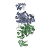
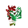
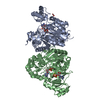
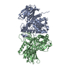
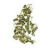
Entry Database : PDB / ID : 5h80Title Biotin Carboxylase domain of single-chain bacterial carboxylase Carboxylase Keywords / / / / / Function / homology Function Domain/homology Component
/ / / / / / / / / / / / / / / / / / / / / / / / / / / / / / / / / / / / / / / / / / Biological species Deinococcus radiodurans (radioresistant)Method / / / Resolution : 1.7 Å Authors Hagmann, A. / Hunkeler, M. / Stuttfeld, E. / Maier, T. Funding support Organization Grant number Country Swiss National Science Foundation 138262 Swiss National Science Foundation 159696 Swiss National Science Foundation 14523
Journal : Structure / Year : 2016Title : Hybrid Structure of a Dynamic Single-Chain Carboxylase from Deinococcus radiodurans.Authors : Hagmann, A. / Hunkeler, M. / Stuttfeld, E. / Maier, T. History Deposition Dec 23, 2015 Deposition site / Processing site Revision 1.0 Jul 20, 2016 Provider / Type Revision 1.1 Aug 17, 2016 Group Revision 1.2 Apr 24, 2019 Group / Source and taxonomy / Category / Item Revision 1.3 Jan 10, 2024 Group / Database references / Refinement descriptionCategory chem_comp_atom / chem_comp_bond ... chem_comp_atom / chem_comp_bond / database_2 / pdbx_initial_refinement_model Item / _database_2.pdbx_database_accession
Show all Show less
 Movie
Movie Controller


 Open data
 Basic information
 Components
 Keywords
 Function and homology information
 Deinococcus radiodurans (radioresistant)
 X-RAY DIFFRACTION /
  Authors
 Switzerland, 3items
  Citation
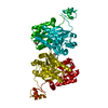
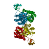
 Structure visualization
 Downloads & links
 Other downloads

 PDBj
PDBj


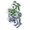
 Assembly
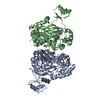

 Spodoptera frugiperda (fall armyworm) / References: UniProt: Q9RYK2
 Mass: 62.068 Da / Num. of mol.: 7 / Source method: obtained synthetically / Formula: C2H6O2
 Mass: 18.015 Da / Num. of mol.: 786 / Source method: isolated from a natural source / Formula: H2O
 Sample preparation
 Processing