 Movie
Movie Controller

[English] 日本語
 Yorodumi
- PDB-2dzd: Crystal structure of the biotin carboxylase domain of pyruvate ca... -
+ Open data

- Basic information
| Entry | Database: PDB / ID: 2dzd | ||||||
|---|---|---|---|---|---|---|---|
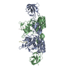
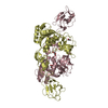
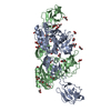
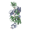
| Title | Crystal structure of the biotin carboxylase domain of pyruvate carboxylase | ||||||
 Components Components | pyruvate carboxylase | ||||||
 Keywords Keywords | LIGASE / biotin carboxylase / pyruvate carboxylase / bacillus thermodenitrificans | ||||||
| Function / homology |  Function and homology information Function and homology informationpyruvate carboxylase activity / gluconeogenesis / ATP binding / metal ion binding / cytoplasm Similarity search - Function | ||||||
| Biological species |  Geobacillus thermodenitrificans (bacteria) Geobacillus thermodenitrificans (bacteria) | ||||||
| Method |  X-RAY DIFFRACTION / SYNCHROTRON / MOLECULAR REPLACEMENT / Resolution: 2.4 Å X-RAY DIFFRACTION / SYNCHROTRON / MOLECULAR REPLACEMENT / Resolution: 2.4 Å | ||||||
 Authors Authors | Kondo, S. / Nakajima, Y. / Sugio, S. / Sueda, S. / Islam, M.N. / Kondo, H. | ||||||
 Citation Citation | Journal: ACTA CRYSTALLOGR.,SECT.D / Year: 2007 Title: Structure of the biotin carboxylase domain of pyruvate carboxylase from Bacillus thermodenitrificans Authors: Kondo, S. / Nakajima, Y. / Sugio, S. / Sueda, S. / Islam, M.N. / Kondo, H. | ||||||
| History |
|
- Structure visualization
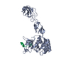
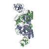
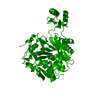
| Structure viewer | Molecule: MolmilJmol/JSmol |
|---|
- Downloads & links
-Download
| PDBx/mmCIF format | 2dzd.cif.gz | 187.8 KB | Display | PDBx/mmCIF format |
|---|---|---|---|---|
| PDB format | pdb2dzd.ent.gz | 150.4 KB | Display | PDB format |
| PDBx/mmJSON format | 2dzd.json.gz | Tree view | PDBx/mmJSON format | |
| Others |  Other downloads Other downloads |
-Validation report
| Arichive directory | https://data.pdbj.org/pub/pdb/validation_reports/dz/2dzdftp://data.pdbj.org/pub/pdb/validation_reports/dz/2dzd | HTTPS FTP |
|---|
-Related structure data
| Related structure data |  1ulzS S: Starting model for refinement |
|---|---|
| Similar structure data |

-Links
 PDBj
PDBj


- Assembly
| Deposited unit | 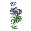
| ||||||||
|---|---|---|---|---|---|---|---|---|---|
| 1 |
| ||||||||
| Unit cell |
| ||||||||
| Details | Unknown, it is not clearly. Maybe the biological assembly part is dimer in asymmetric unit. |
-Components
| #1: Protein | Mass: 51403.406 Da / Num. of mol.: 2 / Fragment: biotin carboxylase domain Source method: isolated from a genetically manipulated source Source: (gene. exp.) Geobacillus thermodenitrificans (bacteria)Plasmid: PTRC99A / Production host: #2: Water | ChemComp-HOH / |  Mass: 18.015 Da / Num. of mol.: 349 / Source method: isolated from a natural source / Formula: H2O Mass: 18.015 Da / Num. of mol.: 349 / Source method: isolated from a natural source / Formula: H2O |
|---|
-Experimental details
-Experiment
| Experiment | Method: X-RAY DIFFRACTION / Number of used crystals: 1 |
|---|
- Sample preparation
| Crystal | Density Matthews: 2.59 Å3/Da / Density % sol: 52.59 % |
|---|---|
| Crystal grow | Temperature: 293 K / Method: vapor diffusion, hanging drop / pH: 7.8 Details: 2% PEG 8000, 100mM Tris, pH 7.8, VAPOR DIFFUSION, HANGING DROP, temperature 293K |
-Data collection
| Diffraction | Mean temperature: 100 K |
|---|---|
| Diffraction source | Source: SYNCHROTRON / Site: SPring-8  / Beamline: BL24XU / Wavelength: 0.836 Å / Beamline: BL24XU / Wavelength: 0.836 Å |
| Detector | Type: RIGAKU JUPITER / Detector: CCD / Date: Nov 6, 2002 |
| Radiation | Protocol: SINGLE WAVELENGTH / Monochromatic (M) / Laue (L): M / Scattering type: x-ray |
| Radiation wavelength | Wavelength: 0.836 Å / Relative weight: 1 |
| Reflection | Resolution: 2.4→20 Å / Num. all: 42671 / Num. obs: 42671 / % possible obs: 100 % / Observed criterion σ(F): 0 / Observed criterion σ(I): 0 / Rsym value: 0.088 |
- Processing
| Software |
| ||||||||||||||||||||
|---|---|---|---|---|---|---|---|---|---|---|---|---|---|---|---|---|---|---|---|---|---|
| Refinement | Method to determine structure: MOLECULAR REPLACEMENT Starting model: 1ULZ Resolution: 2.4→20 Å
| ||||||||||||||||||||
| Refinement step | Cycle: LAST / Resolution: 2.4→20 Å
|
