 Movie
Movie Controller


+ Open data

- Basic information
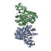
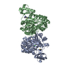
| Entry | Database: PDB / ID: 4h0p | ||||||
|---|---|---|---|---|---|---|---|
| Title | Crystal Structure of Acetate Kinase from Cryptococcus neoformans | ||||||
 Components Components | acetate kinase | ||||||
 Keywords Keywords | TRANSFERASE / ATP-dependent acetate kinase / ASKHA (acetate and sugar kinase / hsc70 / actin) superfamily / Ribonuclease H-like fold | ||||||
| Function / homology |  Function and homology information Function and homology informationacetate kinase / acetate kinase activity / acetate metabolic process / acetyl-CoA biosynthetic process / magnesium ion binding / ATP binding Similarity search - Function | ||||||
| Biological species |  Cryptococcus neoformans (Cryptococcus neoformans serotype A) Cryptococcus neoformans (Cryptococcus neoformans serotype A) | ||||||
| Method |  X-RAY DIFFRACTION / SYNCHROTRON / MOLECULAR REPLACEMENT / Resolution: 1.894 Å X-RAY DIFFRACTION / SYNCHROTRON / MOLECULAR REPLACEMENT / Resolution: 1.894 Å | ||||||
 Authors Authors | Thaker, T.M. / Iverson, T.M. | ||||||
 Citation Citation | Journal: J.Struct.Biol. / Year: 2013 Title: Crystal structures of acetate kinases from the eukaryotic pathogens Entamoeba histolytica and Cryptococcus neoformans. Authors: Thaker, T.M. / Tanabe, M. / Fowler, M.L. / Preininger, A.M. / Ingram-Smith, C. / Smith, K.S. / Iverson, T.M. | ||||||
| History |
|
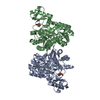
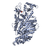
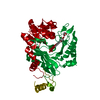
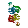
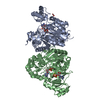
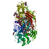
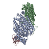
- Structure visualization
| Structure viewer | Molecule: MolmilJmol/JSmol |
|---|
- Downloads & links
-Download
| PDBx/mmCIF format | 4h0p.cif.gz | 180.3 KB | Display | PDBx/mmCIF format |
|---|---|---|---|---|
| PDB format | pdb4h0p.ent.gz | 143.6 KB | Display | PDB format |
| PDBx/mmJSON format | 4h0p.json.gz | Tree view | PDBx/mmJSON format | |
| Others |  Other downloads Other downloads |
-Validation report
| Arichive directory | https://data.pdbj.org/pub/pdb/validation_reports/h0/4h0pftp://data.pdbj.org/pub/pdb/validation_reports/h0/4h0p | HTTPS FTP |
|---|
-Related structure data
| Related structure data | 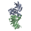 4h0oC 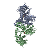 1g99S C: citing same article ( S: Starting model for refinement |
|---|---|
| Similar structure data |

-Links
 PDBj
PDBj- Assembly
| Deposited unit | 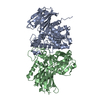
| ||||||||
|---|---|---|---|---|---|---|---|---|---|
| 1 |
| ||||||||
| Unit cell |
|
-Components
| #1: Protein | Mass: 48174.676 Da / Num. of mol.: 2 Source method: isolated from a genetically manipulated source Source: (gene. exp.) Cryptococcus neoformans (Cryptococcus neoformans serotype A)Strain: H99 serotype A / Gene: ACKA / Plasmid: pET21b / Production host:  #2: Water | ChemComp-HOH / |  Mass: 18.015 Da / Num. of mol.: 400 / Source method: isolated from a natural source / Formula: H2O Mass: 18.015 Da / Num. of mol.: 400 / Source method: isolated from a natural source / Formula: H2O |
|---|
-Experimental details
-Experiment
| Experiment | Method: X-RAY DIFFRACTION / Number of used crystals: 1 |
|---|
- Sample preparation
| Crystal | Density Matthews: 2.24 Å3/Da / Density % sol: 44.99 % |
|---|---|
| Crystal grow | Temperature: 277 K / Method: vapor diffusion, hanging drop / pH: 6.6 Details: 18.5% PEG2000, 100 mM sodium tartrate, 50 mM ADA, pH 6.2, VAPOR DIFFUSION, HANGING DROP, temperature 277K |
-Data collection
| Diffraction | Mean temperature: 100 K |
|---|---|
| Diffraction source | Source: SYNCHROTRON / Site: APS  / Beamline: 21-ID-D / Wavelength: 1.12712 Å / Beamline: 21-ID-D / Wavelength: 1.12712 Å |
| Detector | Type: MARMOSAIC 300 mm CCD / Detector: CCD / Date: Jan 31, 2009 |
| Radiation | Monochromator: diamond(111) / Protocol: SINGLE WAVELENGTH / Monochromatic (M) / Laue (L): M / Scattering type: x-ray |
| Radiation wavelength | Wavelength: 1.12712 Å / Relative weight: 1 |
| Reflection | Resolution: 1.89→77.95 Å / Num. all: 67072 / Num. obs: 64297 / % possible obs: 95.9 % / Observed criterion σ(F): 2 / Observed criterion σ(I): 2 / Redundancy: 3.5 % / Rsym value: 0.058 / Net I/σ(I): 27 |
| Reflection shell | Resolution: 1.89→1.97 Å / Redundancy: 3 % / Mean I/σ(I) obs: 5.2 / Rsym value: 0.309 / % possible all: 89.8 |
- Processing
| Software |
| ||||||||||||||||||||||||||||||||||||||||||||||||||||||||||||||||||||||||||||||||||||||||||||||||||||||||||||||||||||||||||||||||||||||||||||||||||||||||||||||||||||||||
|---|---|---|---|---|---|---|---|---|---|---|---|---|---|---|---|---|---|---|---|---|---|---|---|---|---|---|---|---|---|---|---|---|---|---|---|---|---|---|---|---|---|---|---|---|---|---|---|---|---|---|---|---|---|---|---|---|---|---|---|---|---|---|---|---|---|---|---|---|---|---|---|---|---|---|---|---|---|---|---|---|---|---|---|---|---|---|---|---|---|---|---|---|---|---|---|---|---|---|---|---|---|---|---|---|---|---|---|---|---|---|---|---|---|---|---|---|---|---|---|---|---|---|---|---|---|---|---|---|---|---|---|---|---|---|---|---|---|---|---|---|---|---|---|---|---|---|---|---|---|---|---|---|---|---|---|---|---|---|---|---|---|---|---|---|---|---|---|---|---|
| Refinement | Method to determine structure: MOLECULAR REPLACEMENT Starting model: PDB ENTRY 1G99 Resolution: 1.894→38.973 Å / SU ML: 0.26 / σ(F): 1.34 / Phase error: 24.39 / Stereochemistry target values: ML
| ||||||||||||||||||||||||||||||||||||||||||||||||||||||||||||||||||||||||||||||||||||||||||||||||||||||||||||||||||||||||||||||||||||||||||||||||||||||||||||||||||||||||
| Solvent computation | Shrinkage radii: 0.9 Å / VDW probe radii: 1.11 Å / Solvent model: FLAT BULK SOLVENT MODEL | ||||||||||||||||||||||||||||||||||||||||||||||||||||||||||||||||||||||||||||||||||||||||||||||||||||||||||||||||||||||||||||||||||||||||||||||||||||||||||||||||||||||||
| Refinement step | Cycle: LAST / Resolution: 1.894→38.973 Å
| ||||||||||||||||||||||||||||||||||||||||||||||||||||||||||||||||||||||||||||||||||||||||||||||||||||||||||||||||||||||||||||||||||||||||||||||||||||||||||||||||||||||||
| Refine LS restraints |
| ||||||||||||||||||||||||||||||||||||||||||||||||||||||||||||||||||||||||||||||||||||||||||||||||||||||||||||||||||||||||||||||||||||||||||||||||||||||||||||||||||||||||
| LS refinement shell |
|
