 Movie
Movie Controller

[English] 日本語
 Yorodumi
- PDB-5h6z: Crystal structure of Set7, a novel histone methyltransferase in S... -
+ Open data

- Basic information
| Entry | Database: PDB / ID: 5h6z | |||||||||||||||
|---|---|---|---|---|---|---|---|---|---|---|---|---|---|---|---|---|
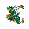
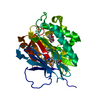
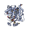
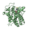
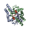
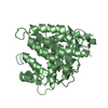
| Title | Crystal structure of Set7, a novel histone methyltransferase in Schizossacharomyces pombe | |||||||||||||||
 Components Components | SET domain-containing protein 7 | |||||||||||||||
 Keywords Keywords | TRANSCRIPTION / Histone / methytransferase / Schizosaccharomyces pombe SET7 / Epigenetic | |||||||||||||||
| Function / homology |  Function and homology information Function and homology informationhistone H3K37 methyltransferase activity / sporulation resulting in formation of a cellular spore / Transferases; Transferring one-carbon groups; Methyltransferases / chromatin organization / methylation / chromatin / nucleus / cytoplasm / cytosol Similarity search - Function | |||||||||||||||
| Biological species |  | |||||||||||||||
| Method |  X-RAY DIFFRACTION / SYNCHROTRON / MOLECULAR REPLACEMENT / Resolution: 2 Å X-RAY DIFFRACTION / SYNCHROTRON / MOLECULAR REPLACEMENT / Resolution: 2 Å | |||||||||||||||
 Authors Authors | Mevius, D.E.H.F. / Shen, Y. / Morishita, M. / Carrozzini, B. / Caliandro, R. / di Luccio, E. | |||||||||||||||
| Funding support |  Korea, Republic Of, Korea, Republic Of,  Italy, 4items Italy, 4items
| |||||||||||||||
 Citation Citation | Journal: Structure / Year: 2019 Title: Set7 Is a H3K37 Methyltransferase in Schizosaccharomyces pombe and Is Required for Proper Gametogenesis. Authors: Shen, Y. / Mevius, D.E.H.F. / Caliandro, R. / Carrozzini, B. / Roh, Y. / Kim, J. / Kim, S. / Ha, S.C. / Morishita, M. / di Luccio, E. | |||||||||||||||
| History |
|
- Structure visualization
| Structure viewer | Molecule: MolmilJmol/JSmol |
|---|
- Downloads & links
-Download
| PDBx/mmCIF format | 5h6z.cif.gz | 73 KB | Display | PDBx/mmCIF format |
|---|---|---|---|---|
| PDB format | pdb5h6z.ent.gz | 52.5 KB | Display | PDB format |
| PDBx/mmJSON format | 5h6z.json.gz | Tree view | PDBx/mmJSON format | |
| Others |  Other downloads Other downloads |
-Validation report
| Arichive directory | https://data.pdbj.org/pub/pdb/validation_reports/h6/5h6zftp://data.pdbj.org/pub/pdb/validation_reports/h6/5h6z | HTTPS FTP |
|---|
-Related structure data
| Related structure data |  5ww0C  3kmaS S: Starting model for refinement C: citing same article ( |
|---|---|
| Similar structure data |
-Links
 PDBj
PDBj

- Assembly
| Deposited unit | 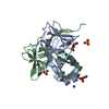
| |||||||||
|---|---|---|---|---|---|---|---|---|---|---|
| 1 |
| |||||||||
| Unit cell |
| |||||||||
| Components on special symmetry positions |
|
-Components
| #1: Protein | Mass: 19597.812 Da / Num. of mol.: 2 Source method: isolated from a genetically manipulated source Source: (gene. exp.) Strain: 972 / ATCC 24843 / Gene: set7, SPCC297.04c / Plasmid: pTYB12 / Production host:  References: UniProt: Q9Y7Q6, histone-lysine N-methyltransferase #2: Chemical |   Mass: 96.063 Da / Num. of mol.: 3 / Source method: obtained synthetically / Formula: SO4 Mass: 96.063 Da / Num. of mol.: 3 / Source method: obtained synthetically / Formula: SO4#3: Chemical | ChemComp-NH4 /   Mass: 18.038 Da / Num. of mol.: 7 / Source method: obtained synthetically / Formula: H4N Mass: 18.038 Da / Num. of mol.: 7 / Source method: obtained synthetically / Formula: H4N#4: Chemical | ChemComp-PEG / |   Mass: 106.120 Da / Num. of mol.: 1 / Source method: isolated from a natural source / Formula: C4H10O3 Mass: 106.120 Da / Num. of mol.: 1 / Source method: isolated from a natural source / Formula: C4H10O3#5: Water | ChemComp-HOH / |  Mass: 18.015 Da / Num. of mol.: 204 / Source method: isolated from a natural source / Formula: H2O Mass: 18.015 Da / Num. of mol.: 204 / Source method: isolated from a natural source / Formula: H2OSequence details | AUTHORS STATE THAT THE RESIDUES, A-5-A0, A148-A151, B-5-B0 AND B148-B151 SHOULD BE LABELED AS ...AUTHORS STATE THAT THE RESIDUES, A-5-A0, A148-A151, B-5-B0 AND B148-B151 SHOULD BE LABELED AS 'PTYB12 MULTIPLE CLONING SITE RESIDUE'. THE RESIDUES, A152-A163 AND B152-A163 SHOULD BE LABELED AS 'HUMAN INFLUENZA HEMAGGLUTI | |
|---|
-Experimental details
-Experiment
| Experiment | Method: X-RAY DIFFRACTION / Number of used crystals: 1 |
|---|
- Sample preparation
| Crystal | Density Matthews: 2.08 Å3/Da / Density % sol: 40.81 % Description: THE ENTRY CONTAINS FRIEDEL PAIRS IN F_PLUS/MINUS COLUMNS. |
|---|---|
| Crystal grow | Temperature: 286 K / Method: vapor diffusion, hanging drop / pH: 7.5 Details: 250mM Ammonium Sulfate, 14% PEG 3.350, 1(protein):1(buffer):0.4 ratio of 0.3M Glycyl-glycyl-glycine (~48mM final conc), Protein concentration(4mg/mL), Protein buffer(10mM Tris pH 7.5) ...Details: 250mM Ammonium Sulfate, 14% PEG 3.350, 1(protein):1(buffer):0.4 ratio of 0.3M Glycyl-glycyl-glycine (~48mM final conc), Protein concentration(4mg/mL), Protein buffer(10mM Tris pH 7.5) Temperature(13), Well volume(500ul), 5uL of HCL (12.1M) was added to the reservoir drop prior to drop setup Temp details: 13 |
-Data collection
| Diffraction | Mean temperature: 100 K |
|---|---|
| Diffraction source | Source: SYNCHROTRON / Site: PAL/PLS / Beamline: 7A (6B, 6C1) / Wavelength: 0.97933 Å |
| Detector | Type: ADSC QUANTUM 270 / Detector: CCD / Date: May 16, 2016 |
| Radiation | Monochromator: DCM Si (111) Crystal / Protocol: SINGLE WAVELENGTH / Monochromatic (M) / Laue (L): M / Scattering type: x-ray |
| Radiation wavelength | Wavelength: 0.97933 Å / Relative weight: 1 |
| Reflection | Resolution: 1.999→57.314 Å / Num. obs: 21580 / % possible obs: 91 % / Redundancy: 5.8 % / Net I/σ(I): 4.2 |
| Reflection shell | Resolution: 2→2.03 Å / Redundancy: 5.8 % / Rmerge(I) obs: 0.806 / Mean I/σ(I) obs: 2.03 / % possible all: 96.4 |
- Processing
| Software |
| ||||||||||||||||||||||||||||||||||||||||||||||||||||||||||||||||||||||||||||||||||||||||||||||||||||||||||||||||||||||||||||||||||||||||||||||||||||||||||||||||||||||||||||||||||||||
|---|---|---|---|---|---|---|---|---|---|---|---|---|---|---|---|---|---|---|---|---|---|---|---|---|---|---|---|---|---|---|---|---|---|---|---|---|---|---|---|---|---|---|---|---|---|---|---|---|---|---|---|---|---|---|---|---|---|---|---|---|---|---|---|---|---|---|---|---|---|---|---|---|---|---|---|---|---|---|---|---|---|---|---|---|---|---|---|---|---|---|---|---|---|---|---|---|---|---|---|---|---|---|---|---|---|---|---|---|---|---|---|---|---|---|---|---|---|---|---|---|---|---|---|---|---|---|---|---|---|---|---|---|---|---|---|---|---|---|---|---|---|---|---|---|---|---|---|---|---|---|---|---|---|---|---|---|---|---|---|---|---|---|---|---|---|---|---|---|---|---|---|---|---|---|---|---|---|---|---|---|---|---|---|
| Refinement | Method to determine structure: MOLECULAR REPLACEMENT Starting model: 3KMA Resolution: 2→57.31 Å / Cor.coef. Fo:Fc: 0.959 / Cor.coef. Fo:Fc free: 0.933 / SU B: 4.134 / SU ML: 0.111 / Cross valid method: THROUGHOUT / ESU R: 0.16 / ESU R Free: 0.154 Details: HYDROGENS HAVE BEEN ADDED IN THE RIDING POSITIONS, SF FILE CONTAINS FRIEDEL PAIRS UNDER I/F_MINUS AND I/ F_PLUS COLUMNS.
| ||||||||||||||||||||||||||||||||||||||||||||||||||||||||||||||||||||||||||||||||||||||||||||||||||||||||||||||||||||||||||||||||||||||||||||||||||||||||||||||||||||||||||||||||||||||
| Solvent computation | Ion probe radii: 0.8 Å / Shrinkage radii: 0.8 Å / VDW probe radii: 1.2 Å | ||||||||||||||||||||||||||||||||||||||||||||||||||||||||||||||||||||||||||||||||||||||||||||||||||||||||||||||||||||||||||||||||||||||||||||||||||||||||||||||||||||||||||||||||||||||
| Displacement parameters | Biso mean: 32.42 Å2
| ||||||||||||||||||||||||||||||||||||||||||||||||||||||||||||||||||||||||||||||||||||||||||||||||||||||||||||||||||||||||||||||||||||||||||||||||||||||||||||||||||||||||||||||||||||||
| Refinement step | Cycle: LAST / Resolution: 2→57.31 Å
| ||||||||||||||||||||||||||||||||||||||||||||||||||||||||||||||||||||||||||||||||||||||||||||||||||||||||||||||||||||||||||||||||||||||||||||||||||||||||||||||||||||||||||||||||||||||
| Refine LS restraints |
|