 Movie
Movie Controller

[English] 日本語
 Yorodumi
- PDB-5gtb: crystal structure of intermembrane space region of the ARC6-PDV2 ... -
+ Open data

- Basic information
| Entry | Database: PDB / ID: 5gtb | ||||||
|---|---|---|---|---|---|---|---|
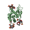
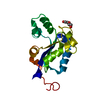
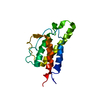
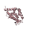
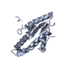
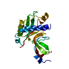
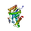
| Title | crystal structure of intermembrane space region of the ARC6-PDV2 complex | ||||||
 Components Components |
| ||||||
 Keywords Keywords | PLANT PROTEIN / beta barrel / key in the lock / complex | ||||||
| Function / homology |  Function and homology information Function and homology informationchloroplast fission / response to gibberellin / chloroplast inner membrane / chloroplast organization / chloroplast outer membrane / chloroplast envelope / phosphatidylinositol-4-phosphate binding / plastid / chloroplast / protein homodimerization activity / mitochondrion Similarity search - Function | ||||||
| Biological species |  | ||||||
| Method |  X-RAY DIFFRACTION / SYNCHROTRON / MOLECULAR REPLACEMENT / Resolution: 2.871 Å X-RAY DIFFRACTION / SYNCHROTRON / MOLECULAR REPLACEMENT / Resolution: 2.871 Å | ||||||
 Authors Authors | Feng, Y. / Wang, W. | ||||||
 Citation Citation | Journal: Nat Plants / Year: 2017 Title: Structural insights into the coordination of plastid division by the ARC6-PDV2 complex Authors: Wang, W. / Li, J. / Sun, Q. / Yu, X. / Zhang, W. / Jia, N. / An, C. / Li, Y. / Dong, Y. / Han, F. / Chang, N. / Liu, X. / Zhu, Z. / Yu, Y. / Fan, S. / Yang, M. / Luo, S.Z. / Gao, H. / Feng, Y. | ||||||
| History |
|
- Structure visualization
| Structure viewer | Molecule: MolmilJmol/JSmol |
|---|
- Downloads & links
-Download
| PDBx/mmCIF format | 5gtb.cif.gz | 44.3 KB | Display | PDBx/mmCIF format |
|---|---|---|---|---|
| PDB format | pdb5gtb.ent.gz | 30 KB | Display | PDB format |
| PDBx/mmJSON format | 5gtb.json.gz | Tree view | PDBx/mmJSON format | |
| Others |  Other downloads Other downloads |
-Validation report
| Arichive directory | https://data.pdbj.org/pub/pdb/validation_reports/gt/5gtbftp://data.pdbj.org/pub/pdb/validation_reports/gt/5gtb | HTTPS FTP |
|---|
-Related structure data
| Related structure data |  5hadSC S: Starting model for refinement C: citing same article ( |
|---|---|
| Similar structure data |


-Links
 PDBj
PDBj

- Assembly
| Deposited unit | 
| ||||||||
|---|---|---|---|---|---|---|---|---|---|
| 1 |
| ||||||||
| Unit cell |
|
-Components
| #1: Protein | Mass: 17320.494 Da / Num. of mol.: 1 Source method: isolated from a genetically manipulated source Source: (gene. exp.)  |
|---|---|
| #2: Protein/peptide | Mass: 2745.134 Da / Num. of mol.: 1 Source method: isolated from a genetically manipulated source Source: (gene. exp.) |
-Experimental details
-Experiment
| Experiment | Method: X-RAY DIFFRACTION / Number of used crystals: 1 |
|---|
- Sample preparation
| Crystal | Density Matthews: 5.51 Å3/Da / Density % sol: 77.7 % |
|---|---|
| Crystal grow | Temperature: 291 K / Method: vapor diffusion / Details: PEG2000MME, Tris pH6.5 |
-Data collection
| Diffraction | Mean temperature: 100 K |
|---|---|
| Diffraction source | Source: SYNCHROTRON / Site: SSRF  / Beamline: BL17U / Wavelength: 0.9796 Å / Beamline: BL17U / Wavelength: 0.9796 Å |
| Detector | Type: ADSC QUANTUM 315r / Detector: CCD / Date: Nov 9, 2015 |
| Radiation | Protocol: SINGLE WAVELENGTH / Monochromatic (M) / Laue (L): M / Scattering type: x-ray |
| Radiation wavelength | Wavelength: 0.9796 Å / Relative weight: 1 |
| Reflection | Resolution: 2.871→27.438 Å / Num. obs: 10239 / % possible obs: 93.82 % / Redundancy: 12.2 % / Rmerge(I) obs: 0.076 / Net I/σ(I): 33.8 |
| Reflection shell | Resolution: 2.87→2.92 Å |
- Processing
| Software |
| ||||||||||||||||||||||||||||
|---|---|---|---|---|---|---|---|---|---|---|---|---|---|---|---|---|---|---|---|---|---|---|---|---|---|---|---|---|---|
| Refinement | Method to determine structure: MOLECULAR REPLACEMENT Starting model: 5HAD Resolution: 2.871→27.438 Å / SU ML: 0.4 / Cross valid method: FREE R-VALUE / σ(F): 1.36 / Phase error: 32.8
| ||||||||||||||||||||||||||||
| Solvent computation | Shrinkage radii: 0.9 Å / VDW probe radii: 1.11 Å | ||||||||||||||||||||||||||||
| Refinement step | Cycle: LAST / Resolution: 2.871→27.438 Å
| ||||||||||||||||||||||||||||
| Refine LS restraints |
| ||||||||||||||||||||||||||||
| LS refinement shell |
|
