 Movie
Movie Controller

[English] 日本語
 Yorodumi
- PDB-5gqu: Crystal structure of branching enzyme from Cyanothece sp. ATCC 51142 -
+ Open data

- Basic information
| Entry | Database: PDB / ID: 5gqu | ||||||||||||
|---|---|---|---|---|---|---|---|---|---|---|---|---|---|
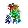
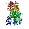
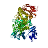
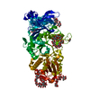
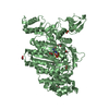
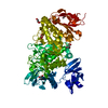
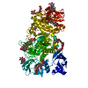
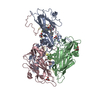
| Title | Crystal structure of branching enzyme from Cyanothece sp. ATCC 51142 | ||||||||||||
 Components Components | 1,4-alpha-glucan branching enzyme GlgB | ||||||||||||
 Keywords Keywords | TRANSFERASE / branching enzyme / glycoside hydrolase family 13 / cyanobacteria / starch | ||||||||||||
| Function / homology |  Function and homology information Function and homology information1,4-alpha-glucan branching enzyme / 1,4-alpha-glucan branching enzyme activity / glycogen biosynthetic process / hydrolase activity, hydrolyzing O-glycosyl compounds / metal ion binding / cytosol Similarity search - Function | ||||||||||||
| Biological species | Cyanothece sp. | ||||||||||||
| Method |  X-RAY DIFFRACTION / SYNCHROTRON / MOLECULAR REPLACEMENT / Resolution: 1.85 Å X-RAY DIFFRACTION / SYNCHROTRON / MOLECULAR REPLACEMENT / Resolution: 1.85 Å | ||||||||||||
 Authors Authors | Suzuki, R. / Suzuki, E. | ||||||||||||
| Funding support |  Japan, 3items Japan, 3items
| ||||||||||||
 Citation Citation | Journal: J. Biol. Chem. / Year: 2017 Title: Bound Substrate in the Structure of Cyanobacterial Branching Enzyme Supports a New Mechanistic Model Authors: Hayashi, M. / Suzuki, R. / Colleoni, C. / Ball, S.G. / Fujita, N. / Suzuki, E. #1: Journal: Acta Crystallogr F Struct Biol Commun / Year: 2015 Title: Crystallization and crystallographic analysis of branching enzymes from Cyanothece sp. ATCC 51142. Authors: Hayashi, M. / Suzuki, R. / Colleoni, C. / Ball, S.G. / Fujita, N. / Suzuki, E. | ||||||||||||
| History |
|
- Structure visualization
| Structure viewer | Molecule: MolmilJmol/JSmol |
|---|
- Downloads & links
-Download
| PDBx/mmCIF format | 5gqu.cif.gz | 194.7 KB | Display | PDBx/mmCIF format |
|---|---|---|---|---|
| PDB format | pdb5gqu.ent.gz | 150.3 KB | Display | PDB format |
| PDBx/mmJSON format | 5gqu.json.gz | Tree view | PDBx/mmJSON format | |
| Others |  Other downloads Other downloads |
-Validation report
| Arichive directory | https://data.pdbj.org/pub/pdb/validation_reports/gq/5gquftp://data.pdbj.org/pub/pdb/validation_reports/gq/5gqu | HTTPS FTP |
|---|
-Related structure data
| Related structure data | 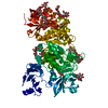 5gqvC 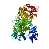 5gqwC  5gqxC 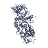 1m7xS C: citing same article ( S: Starting model for refinement |
|---|---|
| Similar structure data |


-Links
 PDBj
PDBj

- Assembly
| Deposited unit | 
| ||||||||
|---|---|---|---|---|---|---|---|---|---|
| 1 |
| ||||||||
| Unit cell |
|
-Components
| #1: Protein | Mass: 92686.422 Da / Num. of mol.: 1 Source method: isolated from a genetically manipulated source Source: (gene. exp.)  Cyanothece sp. (strain ATCC 51142) (bacteria) Cyanothece sp. (strain ATCC 51142) (bacteria)Strain: ATCC 51142 / Gene: glgB, glgB1, cce_2248 / Plasmid: pET15B / Production host: References: UniProt: B1WPM8, 1,4-alpha-glucan branching enzyme | ||||
|---|---|---|---|---|---|
| #2: Chemical | ChemComp-GOL /   Mass: 92.094 Da / Num. of mol.: 7 / Source method: obtained synthetically / Formula: C3H8O3 Mass: 92.094 Da / Num. of mol.: 7 / Source method: obtained synthetically / Formula: C3H8O3#3: Chemical | ChemComp-MG /   Mass: 24.305 Da / Num. of mol.: 4 / Source method: obtained synthetically / Formula: Mg Mass: 24.305 Da / Num. of mol.: 4 / Source method: obtained synthetically / Formula: Mg#4: Water | ChemComp-HOH / |  Mass: 18.015 Da / Num. of mol.: 852 / Source method: isolated from a natural source / Formula: H2O Mass: 18.015 Da / Num. of mol.: 852 / Source method: isolated from a natural source / Formula: H2O |
-Experimental details
-Experiment
| Experiment | Method: X-RAY DIFFRACTION / Number of used crystals: 1 |
|---|
- Sample preparation
| Crystal | Density Matthews: 4.68 Å3/Da / Density % sol: 73.74 % |
|---|---|
| Crystal grow | Temperature: 293 K / Method: vapor diffusion, hanging drop / pH: 7.5 / Details: magnesium chloride, ethanol, HEPES-NaOH / PH range: 7.2 - 7.9 |
-Data collection
| Diffraction | Mean temperature: 100 K |
|---|---|
| Diffraction source | Source: SYNCHROTRON / Site: Photon Factory / Beamline: BL-5A / Wavelength: 1 Å |
| Detector | Type: ADSC QUANTUM 315r / Detector: CCD / Date: Jun 9, 2014 |
| Radiation | Monochromator: Si(111) / Protocol: SINGLE WAVELENGTH / Monochromatic (M) / Laue (L): M / Scattering type: x-ray |
| Radiation wavelength | Wavelength: 1 Å / Relative weight: 1 |
| Reflection | Resolution: 1.85→50 Å / Num. obs: 143511 / % possible obs: 100 % / Redundancy: 14.6 % / Rmerge(I) obs: 0.074 / Net I/σ(I): 61 |
| Reflection shell | Resolution: 1.85→1.88 Å / Redundancy: 14.8 % / Rmerge(I) obs: 0.274 / Mean I/σ(I) obs: 10.7 / % possible all: 100 |
- Processing
| Software |
| ||||||||||||||||||||||||||||||||||||||||||||||||||||||||||||||||||||||||||||||||||||||||||||||||||||||||||||||||||||||||||||||||||||||||||||||||||||||||||||||||||||||||||||||||||||||
|---|---|---|---|---|---|---|---|---|---|---|---|---|---|---|---|---|---|---|---|---|---|---|---|---|---|---|---|---|---|---|---|---|---|---|---|---|---|---|---|---|---|---|---|---|---|---|---|---|---|---|---|---|---|---|---|---|---|---|---|---|---|---|---|---|---|---|---|---|---|---|---|---|---|---|---|---|---|---|---|---|---|---|---|---|---|---|---|---|---|---|---|---|---|---|---|---|---|---|---|---|---|---|---|---|---|---|---|---|---|---|---|---|---|---|---|---|---|---|---|---|---|---|---|---|---|---|---|---|---|---|---|---|---|---|---|---|---|---|---|---|---|---|---|---|---|---|---|---|---|---|---|---|---|---|---|---|---|---|---|---|---|---|---|---|---|---|---|---|---|---|---|---|---|---|---|---|---|---|---|---|---|---|---|
| Refinement | Method to determine structure: MOLECULAR REPLACEMENT Starting model: 1M7X Resolution: 1.85→47.29 Å / Cor.coef. Fo:Fc: 0.971 / Cor.coef. Fo:Fc free: 0.961 / SU B: 1.441 / SU ML: 0.044 / Cross valid method: THROUGHOUT / ESU R: 0.071 / ESU R Free: 0.074 / Stereochemistry target values: MAXIMUM LIKELIHOOD / Details: HYDROGENS HAVE BEEN ADDED IN THE RIDING POSITIONS
| ||||||||||||||||||||||||||||||||||||||||||||||||||||||||||||||||||||||||||||||||||||||||||||||||||||||||||||||||||||||||||||||||||||||||||||||||||||||||||||||||||||||||||||||||||||||
| Solvent computation | Ion probe radii: 0.8 Å / Shrinkage radii: 0.8 Å / VDW probe radii: 1.2 Å / Solvent model: MASK | ||||||||||||||||||||||||||||||||||||||||||||||||||||||||||||||||||||||||||||||||||||||||||||||||||||||||||||||||||||||||||||||||||||||||||||||||||||||||||||||||||||||||||||||||||||||
| Displacement parameters | Biso mean: 24.695 Å2
| ||||||||||||||||||||||||||||||||||||||||||||||||||||||||||||||||||||||||||||||||||||||||||||||||||||||||||||||||||||||||||||||||||||||||||||||||||||||||||||||||||||||||||||||||||||||
| Refinement step | Cycle: LAST / Resolution: 1.85→47.29 Å
| ||||||||||||||||||||||||||||||||||||||||||||||||||||||||||||||||||||||||||||||||||||||||||||||||||||||||||||||||||||||||||||||||||||||||||||||||||||||||||||||||||||||||||||||||||||||
| Refine LS restraints |
|