 Movie
Movie Controller


+ Open data

- Basic information
| Entry | Database: PDB / ID: 5gkj | ||||||||||||
|---|---|---|---|---|---|---|---|---|---|---|---|---|---|
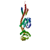
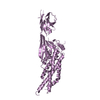
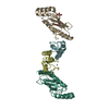
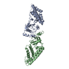
| Title | Structure of EndoMS in apo form | ||||||||||||
 Components Components | Endonuclease EndoMS | ||||||||||||
 Keywords Keywords | HYDROLASE / ENDONUCLEASE / DNA-BINDING | ||||||||||||
| Function / homology |  Function and homology information Function and homology informationsingle-stranded DNA endodeoxyribonuclease activity / Hydrolases; Acting on ester bonds / DNA binding / cytoplasm Similarity search - Function | ||||||||||||
| Biological species |   Thermococcus kodakarensis KOD1 (archaea) Thermococcus kodakarensis KOD1 (archaea) | ||||||||||||
| Method |  X-RAY DIFFRACTION / SYNCHROTRON / MOLECULAR REPLACEMENT / Resolution: 3.2 Å X-RAY DIFFRACTION / SYNCHROTRON / MOLECULAR REPLACEMENT / Resolution: 3.2 Å | ||||||||||||
 Authors Authors | Nakae, S. / Hijikata, A. / Tsuji, T. / Yonezawa, K. / Kouyama, K. / Mayanagi, K. / Ishino, S. / Ishino, Y. / Shirai, T. | ||||||||||||
| Funding support |  Japan, 3items Japan, 3items
| ||||||||||||
 Citation Citation | Journal: Structure / Year: 2016 Title: Structure of the EndoMS-DNA Complex as Mismatch Restriction Endonuclease Authors: Nakae, S. / Hijikata, A. / Tsuji, T. / Yonezawa, K. / Kouyama, K.I. / Mayanagi, K. / Ishino, S. / Ishino, Y. / Shirai, T. | ||||||||||||
| History |
|
- Structure visualization
| Structure viewer | Molecule: MolmilJmol/JSmol |
|---|
- Downloads & links
-Download
| PDBx/mmCIF format | 5gkj.cif.gz | 100.4 KB | Display | PDBx/mmCIF format |
|---|---|---|---|---|
| PDB format | pdb5gkj.ent.gz | 76.2 KB | Display | PDB format |
| PDBx/mmJSON format | 5gkj.json.gz | Tree view | PDBx/mmJSON format | |
| Others |  Other downloads Other downloads |
-Validation report
| Summary document | 5gkj_validation.pdf.gz | 449.3 KB | Display | wwPDB validaton report |
|---|---|---|---|---|
| Full document | 5gkj_full_validation.pdf.gz | 456.5 KB | Display | |
| Data in XML | 5gkj_validation.xml.gz | 17.5 KB | Display | |
| Data in CIF | 5gkj_validation.cif.gz | 22.5 KB | Display | |
| Arichive directory | https://data.pdbj.org/pub/pdb/validation_reports/gk/5gkjftp://data.pdbj.org/pub/pdb/validation_reports/gk/5gkj | HTTPS FTP |
-Related structure data
| Related structure data |  5gkeC  5gkfC 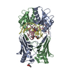 5gkgC  5gkhC  5gkiC  5bsl C: citing same article ( S: Starting model for refinement |
|---|---|
| Similar structure data |


-Links
 PDBj
PDBj- Assembly
| Deposited unit | 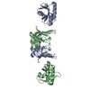
| ||||||||
|---|---|---|---|---|---|---|---|---|---|
| 1 |
| ||||||||
| Unit cell |
|
-Components
| #1: Protein | Mass: 28598.096 Da / Num. of mol.: 2 / Mutation: D165A Source method: isolated from a genetically manipulated source Source: (gene. exp.) Thermococcus kodakarensis KOD1 (archaea)Strain: KOD1 / Gene: TK1898 / Production host:  References: UniProt: Q5JER9, Hydrolases; Acting on ester bonds |
|---|
-Experimental details
-Experiment
| Experiment | Method: X-RAY DIFFRACTION / Number of used crystals: 1 |
|---|
- Sample preparation
| Crystal | Density Matthews: 2.94 Å3/Da / Density % sol: 58.22 % |
|---|---|
| Crystal grow | Temperature: 291 K / Method: vapor diffusion, hanging drop / pH: 6.5 Details: 0.16 M CALCIUM ACETATE, 80 MM SODIUM CACODYLATE, 14.4%(W/V) PEG8000, 20%(W/V) GLYCEROL |
-Data collection
| Diffraction | Mean temperature: 100 K |
|---|---|
| Diffraction source | Source: SYNCHROTRON / Site: Photon Factory / Beamline: BL-17A / Wavelength: 0.98 Å |
| Detector | Type: ADSC QUANTUM 270 / Detector: CCD / Date: May 23, 2014 |
| Radiation | Protocol: SINGLE WAVELENGTH / Monochromatic (M) / Laue (L): M / Scattering type: x-ray |
| Radiation wavelength | Wavelength: 0.98 Å / Relative weight: 1 |
| Reflection | Resolution: 3.2→24.92 Å / Num. obs: 10762 / % possible obs: 97.6 % / Redundancy: 6.8 % / Rmerge(I) obs: 0.073 / Net I/σ(I): 17.7 |
| Reflection shell | Resolution: 3.2→3.37 Å / Redundancy: 7 % / Mean I/σ(I) obs: 10.1 / % possible all: 99.9 |
- Processing
| Software |
| |||||||||||||||||||||||||||||||||||
|---|---|---|---|---|---|---|---|---|---|---|---|---|---|---|---|---|---|---|---|---|---|---|---|---|---|---|---|---|---|---|---|---|---|---|---|---|
| Refinement | Method to determine structure: MOLECULAR REPLACEMENT Starting model: 5BSL 5bsl Resolution: 3.2→24.11 Å / Cross valid method: FREE R-VALUE / σ(F): 1.35 / Phase error: 32.38
| |||||||||||||||||||||||||||||||||||
| Solvent computation | Shrinkage radii: 0.9 Å / VDW probe radii: 1.11 Å | |||||||||||||||||||||||||||||||||||
| Refinement step | Cycle: LAST / Resolution: 3.2→24.11 Å
| |||||||||||||||||||||||||||||||||||
| Refine LS restraints |
| |||||||||||||||||||||||||||||||||||
| LS refinement shell |
|
