 ムービー
ムービー コントローラー


+ データを開く

- 基本情報
基本情報
| 登録情報 | データベース: PDB / ID: 5gjv | ||||||||||||
|---|---|---|---|---|---|---|---|---|---|---|---|---|---|
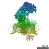
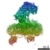
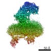
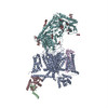
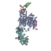
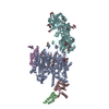
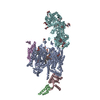
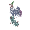
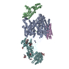
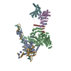
| タイトル | Structure of the mammalian voltage-gated calcium channel Cav1.1 complex at near atomic resolution | ||||||||||||
 要素 要素 |
| ||||||||||||
 キーワード キーワード | MEMBRANE PROTEIN / complex / channel | ||||||||||||
| 機能・相同性 |  機能・相同性情報 機能・相同性情報positive regulation of muscle contraction / high voltage-gated calcium channel activity / L-type voltage-gated calcium channel complex / regulation of calcium ion transmembrane transport via high voltage-gated calcium channel / cellular response to caffeine / calcium channel regulator activity / calcium ion import across plasma membrane / voltage-gated calcium channel activity / release of sequestered calcium ion into cytosol / regulation of ryanodine-sensitive calcium-release channel activity ...positive regulation of muscle contraction / high voltage-gated calcium channel activity / L-type voltage-gated calcium channel complex / regulation of calcium ion transmembrane transport via high voltage-gated calcium channel / cellular response to caffeine / calcium channel regulator activity / calcium ion import across plasma membrane / voltage-gated calcium channel activity / release of sequestered calcium ion into cytosol / regulation of ryanodine-sensitive calcium-release channel activity / T-tubule / muscle contraction / calcium ion transmembrane transport / sarcolemma / transmembrane transporter binding / calmodulin binding / metal ion binding / plasma membrane 類似検索 - 分子機能 | ||||||||||||
| 生物種 |  | ||||||||||||
| 手法 | 電子顕微鏡法 / 単粒子再構成法 / クライオ電子顕微鏡法 / 解像度: 3.6 Å | ||||||||||||
 データ登録者 データ登録者 | Wu, J.P. / Yan, Z. / Li, Z.Q. / Zhou, Q. / Yan, N. | ||||||||||||
| 資金援助 |  中国, 2件 中国, 2件
| ||||||||||||
 引用 引用 | ジャーナル: Nature / 年: 2016 タイトル: Structure of the voltage-gated calcium channel Ca(v)1.1 at 3.6 Å resolution. 著者: Jianping Wu / Zhen Yan / Zhangqiang Li / Xingyang Qian / Shan Lu / Mengqiu Dong / Qiang Zhou / Nieng Yan / 要旨: The voltage-gated calcium (Ca) channels convert membrane electrical signals to intracellular Ca-mediated events. Among the ten subtypes of Ca channel in mammals, Ca1.1 is specified for the excitation- ...The voltage-gated calcium (Ca) channels convert membrane electrical signals to intracellular Ca-mediated events. Among the ten subtypes of Ca channel in mammals, Ca1.1 is specified for the excitation-contraction coupling of skeletal muscles. Here we present the cryo-electron microscopy structure of the rabbit Ca1.1 complex at a nominal resolution of 3.6 Å. The inner gate of the ion-conducting α1-subunit is closed and all four voltage-sensing domains adopt an 'up' conformation, suggesting a potentially inactivated state. The extended extracellular loops of the pore domain, which are stabilized by multiple disulfide bonds, form a windowed dome above the selectivity filter. One side of the dome provides the docking site for the α2δ-1-subunit, while the other side may attract cations through its negative surface potential. The intracellular I-II and III-IV linker helices interact with the β-subunit and the carboxy-terminal domain of α1, respectively. Classification of the particles yielded two additional reconstructions that reveal pronounced displacement of β and adjacent elements in α1. The atomic model of the Ca1.1 complex establishes a foundation for mechanistic understanding of excitation-contraction coupling and provides a three-dimensional template for molecular interpretations of the functions and disease mechanisms of Ca and Na channels. | ||||||||||||
| 履歴 |
|
- 構造の表示
構造の表示
| ムービー |
ムービービューア |
|---|---|
| 構造ビューア | 分子: MolmilJmol/JSmol |
- ダウンロードとリンク
-ダウンロード
| PDBx/mmCIF形式 | 5gjv.cif.gz | 585 KB | 表示 | PDBx/mmCIF形式 |
|---|---|---|---|---|
| PDB形式 | pdb5gjv.ent.gz | 457.8 KB | 表示 | PDB形式 |
| PDBx/mmJSON形式 | 5gjv.json.gz | ツリー表示 | PDBx/mmJSON形式 | |
| その他 |  その他のダウンロード その他のダウンロード |
-検証レポート
| 文書・要旨 | 5gjv_validation.pdf.gz | 1.7 MB | 表示 | wwPDB検証レポート |
|---|---|---|---|---|
| 文書・詳細版 | 5gjv_full_validation.pdf.gz | 1.8 MB | 表示 | |
| XML形式データ | 5gjv_validation.xml.gz | 88.8 KB | 表示 | |
| CIF形式データ | 5gjv_validation.cif.gz | 129.4 KB | 表示 | |
| アーカイブディレクトリ | https://data.pdbj.org/pub/pdb/validation_reports/gj/5gjvftp://data.pdbj.org/pub/pdb/validation_reports/gj/5gjv | HTTPS FTP |
-関連構造データ


-リンク
 PDBj
PDBj


- 集合体
集合体
| 登録構造単位 | 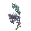
|
|---|---|
| 1 |
|
-要素
-タンパク質 , 1種, 1分子 A
| #1: タンパク質 | 分子量: 212240.594 Da / 分子数: 1 / 由来タイプ: 天然 / 由来: (天然) |
|---|
-Voltage-dependent L-type calcium channel subunit beta- ... , 2種, 2分子 BC
| #2: タンパク質 | 分子量: 11974.729 Da / 分子数: 1 / Fragment: UNP residues 80-174 / 由来タイプ: 組換発現 詳細: This domain was docked by a crystal structure (4DEY) 由来: (組換発現)  |
|---|---|
| #3: タンパク質 | 分子量: 22356.846 Da / 分子数: 1 / Fragment: UNP residues 265-463 / 由来タイプ: 組換発現 詳細: This domain was docked by a crystal structure (4DEY) 由来: (組換発現) |
-Voltage-dependent calcium channel ... , 2種, 2分子 EF
| #4: タンパク質 | 分子量: 25082.254 Da / 分子数: 1 / 由来タイプ: 天然 / 由来: (天然) |
|---|---|
| #5: タンパク質 | 分子量: 125156.000 Da / 分子数: 1 / 由来タイプ: 天然 / 由来: (天然) |
-糖 , 4種, 15分子 
| #6: 多糖 | 2-acetamido-2-deoxy-beta-D-glucopyranose-(1-4)-2-acetamido-2-deoxy-beta-D-glucopyranose #7: 多糖 | #8: 多糖 | 2-acetamido-2-deoxy-beta-D-glucopyranose-(1-4)-2-acetamido-2-deoxy-beta-D-glucopyranose-(1-4)-2- ...2-acetamido-2-deoxy-beta-D-glucopyranose-(1-4)-2-acetamido-2-deoxy-beta-D-glucopyranose-(1-4)-2-acetamido-2-deoxy-beta-D-glucopyranose / triacetyl-beta-chitotriose |   #9: 糖 | ChemComp-NAG /  タイプ: D-saccharide, beta linking / 分子量: 221.208 Da / 分子数: 8 / 由来タイプ: 組換発現 / 式: C8H15NO6 タイプ: D-saccharide, beta linking / 分子量: 221.208 Da / 分子数: 8 / 由来タイプ: 組換発現 / 式: C8H15NO6 |
|---|
-非ポリマー , 2種, 17分子 

| #10: 化合物 | ChemComp-PC1 /  分子量: 790.145 Da / 分子数: 14 / 由来タイプ: 合成 / 式: C44H88NO8P / コメント: リン脂質*YM 分子量: 790.145 Da / 分子数: 14 / 由来タイプ: 合成 / 式: C44H88NO8P / コメント: リン脂質*YM#11: 化合物 |  分子量: 40.078 Da / 分子数: 3 / 由来タイプ: 合成 / 式: Ca 分子量: 40.078 Da / 分子数: 3 / 由来タイプ: 合成 / 式: Ca |
|---|
-詳細
| 配列の詳細 | RESIDUE F1075 IS DUE TO GPI MODIFICATI |
|---|
-実験情報
-実験
| 実験 | 手法: 電子顕微鏡法 |
|---|---|
| EM実験 | 試料の集合状態: PARTICLE / 3次元再構成法: 単粒子再構成法 |
- 試料調製
試料調製
| 構成要素 | 名称: voltage-gated calcium channel Cav1.1 / タイプ: COMPLEX 詳細: The protein complex was endogenous purified from rabbit muscle tissue. Entity ID: #1-#5 / 由来: NATURAL |
|---|---|
| 分子量 | 値: 0.5 MDa / 実験値: NO |
| 由来(天然) | 生物種: |
| 緩衝液 | pH: 8 詳細: 100 mM Tris-HCl, pH 8.0,200 mM NaCl, 10 mM CaCl2,15 mM reduced glutathione, 0.1% digitonin, and protease inhibitors. |
| 試料 | 濃度: 2 mg/ml / 包埋: NO / シャドウイング: NO / 染色: NO / 凍結: YES / 詳細: This sample was monodisperse. |
| 試料支持 | グリッドの材料: COPPER / グリッドのサイズ: 300 divisions/in. / グリッドのタイプ: Quantifoil R1.2/1.3 |
| 急速凍結 | 装置: FEI VITROBOT MARK IV / 凍結剤: ETHANE / 湿度: 100 % / 凍結前の試料温度: 281 K 詳細: Grids were blotted for 2 s and flash-frozen in liquid ethane cooled by liquid nitrogen |
- 電子顕微鏡撮影
| 実験機器 |  モデル: Titan Krios / 画像提供: FEI Company |
|---|---|
| 顕微鏡 | モデル: FEI TITAN KRIOS |
| 電子銃 | 電子線源:  FIELD EMISSION GUN / 加速電圧: 300 kV / 照射モード: SPOT SCAN FIELD EMISSION GUN / 加速電圧: 300 kV / 照射モード: SPOT SCAN |
| 電子レンズ | モード: BRIGHT FIELD / Calibrated defocus min: 1.3 nm / 最大 デフォーカス(補正後): 2.9 nm / アライメント法: COMA FREE |
| 試料ホルダ | 凍結剤: NITROGEN 試料ホルダーモデル: FEI TITAN KRIOS AUTOGRID HOLDER 最低温度: 70 K |
| 撮影 | 平均露光時間: 8 sec. / 電子線照射量: 50 e/Å2 / 検出モード: SUPER-RESOLUTION フィルム・検出器のモデル: GATAN K2 SUMMIT (4k x 4k) 撮影したグリッド数: 5 / 実像数: 9704 |
| 画像スキャン | 動画フレーム数/画像: 32 |
- 解析
解析
| EMソフトウェア |
| ||||||||||||||||||||||||||||||||||||||||||||
|---|---|---|---|---|---|---|---|---|---|---|---|---|---|---|---|---|---|---|---|---|---|---|---|---|---|---|---|---|---|---|---|---|---|---|---|---|---|---|---|---|---|---|---|---|---|
| CTF補正 | タイプ: PHASE FLIPPING AND AMPLITUDE CORRECTION | ||||||||||||||||||||||||||||||||||||||||||||
| 粒子像の選択 | 選択した粒子像数: 1630270 | ||||||||||||||||||||||||||||||||||||||||||||
| 対称性 | 点対称性: C1 (非対称) | ||||||||||||||||||||||||||||||||||||||||||||
| 3次元再構成 | 解像度: 3.6 Å / 解像度の算出法: FSC 0.143 CUT-OFF / 粒子像の数: 527833 / 対称性のタイプ: POINT | ||||||||||||||||||||||||||||||||||||||||||||
| 原子モデル構築 | プロトコル: RIGID BODY FIT | ||||||||||||||||||||||||||||||||||||||||||||
| 精密化 | 最高解像度: 3.6 Å |
