 Movie
Movie Controller

[English] 日本語
 Yorodumi
- PDB-5g5o: Structure of the snake adenovirus 1 hexon-interlacing LH3 protein... -
+ Open data

- Basic information
| Entry | Database: PDB / ID: 5g5o | ||||||
|---|---|---|---|---|---|---|---|
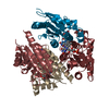
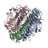
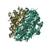
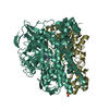
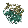
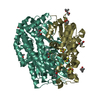
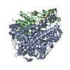
| Title | Structure of the snake adenovirus 1 hexon-interlacing LH3 protein, native | ||||||
 Components Components | LH3 HEXON-INTERLACING CAPSID PROTEIN | ||||||
 Keywords Keywords | VIRAL PROTEIN / CAPSID PROTEIN / BETA-HELIX | ||||||
| Function / homology | Pectin lyase fold / Pectin lyase fold/virulence factor / virion component / ACETATE ION / Hexon-interlacing protein LH3 Function and homology information Function and homology information | ||||||
| Biological species |  SNAKE ADENOVIRUS 1 SNAKE ADENOVIRUS 1 | ||||||
| Method |  X-RAY DIFFRACTION / SYNCHROTRON / MOLECULAR REPLACEMENT / Resolution: 2 Å X-RAY DIFFRACTION / SYNCHROTRON / MOLECULAR REPLACEMENT / Resolution: 2 Å | ||||||
 Authors Authors | Nguyen, T.H. / Singh, A.K. / Albala-Perez, B. / van Raaij, M.J. | ||||||
 Citation Citation | Journal: Structure / Year: 2017 Title: Structure of a Reptilian Adenovirus Reveals a Phage Tailspike Fold Stabilizing a Vertebrate Virus Capsid. Authors: Rosa Menéndez-Conejero / Thanh H Nguyen / Abhimanyu K Singh / Gabriela N Condezo / Rachel E Marschang / Mark J van Raaij / Carmen San Martín /    Abstract: Although non-human adenoviruses (AdVs) might offer solutions to problems posed by human AdVs as therapeutic vectors, little is known about their basic biology. In particular, there are no structural ...Although non-human adenoviruses (AdVs) might offer solutions to problems posed by human AdVs as therapeutic vectors, little is known about their basic biology. In particular, there are no structural studies on the complete virion of any AdV with a non-mammalian host. We combine mass spectrometry, cryo-electron microscopy, and protein crystallography to characterize the composition and structure of a snake AdV (SnAdV-1, Atadenovirus genus). SnAdV-1 particles contain the genus-specific proteins LH3, p32k, and LH2, a previously unrecognized structural component. Remarkably, the cementing protein LH3 has a trimeric β helix fold typical of bacteriophage host attachment proteins. The organization of minor coat proteins differs from that in human AdVs, correlating with higher thermostability in SnAdV-1. These findings add a new piece to the intriguing puzzle of virus evolution, hint at the use of cell entry pathways different from those in human AdVs, and will help development of new, thermostable SnAdV-1-based vectors. | ||||||
| History |
|
- Structure visualization
| Structure viewer | Molecule: MolmilJmol/JSmol |
|---|
- Downloads & links
-Download
| PDBx/mmCIF format | 5g5o.cif.gz | 423 KB | Display | PDBx/mmCIF format |
|---|---|---|---|---|
| PDB format | pdb5g5o.ent.gz | 343.8 KB | Display | PDB format |
| PDBx/mmJSON format | 5g5o.json.gz | Tree view | PDBx/mmJSON format | |
| Others |  Other downloads Other downloads |
-Validation report
| Arichive directory | https://data.pdbj.org/pub/pdb/validation_reports/g5/5g5oftp://data.pdbj.org/pub/pdb/validation_reports/g5/5g5o | HTTPS FTP |
|---|
-Related structure data
| Related structure data |  3599C  5g5nSC S: Starting model for refinement C: citing same article ( |
|---|---|
| Similar structure data |

-Links
 PDBj
PDBj- Assembly
| Deposited unit | 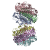
| ||||||||
|---|---|---|---|---|---|---|---|---|---|
| 1 | 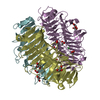
| ||||||||
| 2 | 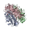
| ||||||||
| Unit cell |
|
-Components
| #1: Protein | Mass: 40241.266 Da / Num. of mol.: 6 Source method: isolated from a genetically manipulated source Source: (gene. exp.) SNAKE ADENOVIRUS 1 / Production host:  #2: Chemical | ChemComp-ACT /   Mass: 59.044 Da / Num. of mol.: 11 / Source method: obtained synthetically / Formula: C2H3O2 Mass: 59.044 Da / Num. of mol.: 11 / Source method: obtained synthetically / Formula: C2H3O2#3: Chemical | ChemComp-GOL /   Mass: 92.094 Da / Num. of mol.: 8 / Source method: obtained synthetically / Formula: C3H8O3 Mass: 92.094 Da / Num. of mol.: 8 / Source method: obtained synthetically / Formula: C3H8O3#4: Chemical | ChemComp-CL /   Mass: 35.453 Da / Num. of mol.: 6 / Source method: obtained synthetically / Formula: Cl Mass: 35.453 Da / Num. of mol.: 6 / Source method: obtained synthetically / Formula: Cl#5: Water | ChemComp-HOH / |  Mass: 18.015 Da / Num. of mol.: 1525 / Source method: isolated from a natural source / Formula: H2O Mass: 18.015 Da / Num. of mol.: 1525 / Source method: isolated from a natural source / Formula: H2OSequence details | WE FOUND SEVERAL SEQUENCE DIFFERENCES BETWEEN OUR PCR- AMPLIFIED GENE AND THE DATABASE. HOWEVER, WE ...WE FOUND SEVERAL SEQUENCE DIFFERENCE | |
|---|
-Experimental details
-Experiment
| Experiment | Method: X-RAY DIFFRACTION / Number of used crystals: 1 |
|---|
- Sample preparation
| Crystal | Density Matthews: 2.13 Å3/Da / Density % sol: 42.3 % / Description: NONE |
|---|---|
| Crystal grow | pH: 8.5 Details: 10 MM TRIS-HCL PH 8.5, 20% (W/V) PEG3350, 0.2 M AMMONIUM ACETATE |
-Data collection
| Diffraction | Mean temperature: 100 K |
|---|---|
| Diffraction source | Source: SYNCHROTRON / Site: ESRF  / Beamline: BM30A / Wavelength: 0.9797 / Beamline: BM30A / Wavelength: 0.9797 |
| Detector | Type: ADSC QUANTUM 315r / Detector: CCD / Date: Jul 11, 2013 Details: TWO CYLINDRICAL PARABOLIC VERTICAL FOCUSSING MIRRORS |
| Radiation | Monochromator: DOUBLE SI(111) CRYSTAL FIXED-EXIT BRAGG MONOCHROMATOR Protocol: SINGLE WAVELENGTH / Monochromatic (M) / Laue (L): M / Scattering type: x-ray |
| Radiation wavelength | Wavelength: 0.9797 Å / Relative weight: 1 |
| Reflection | Resolution: 2→30 Å / Num. obs: 130350 / % possible obs: 94.3 % / Redundancy: 3.3 % / Rmerge(I) obs: 0.2 / Net I/σ(I): 3.8 |
| Reflection shell | Resolution: 2→2.11 Å / Redundancy: 3.4 % / Rmerge(I) obs: 0.72 / Mean I/σ(I) obs: 1.4 / % possible all: 87.6 |
- Processing
| Software |
| ||||||||||||||||||||||||||||||||||||||||||||||||||||||||||||||||||||||||||||||||||||||||||||||||||||||||||||||||||||||||||||||||||||||||||||||||||||||||||||||||||||||||||||||||||||||
|---|---|---|---|---|---|---|---|---|---|---|---|---|---|---|---|---|---|---|---|---|---|---|---|---|---|---|---|---|---|---|---|---|---|---|---|---|---|---|---|---|---|---|---|---|---|---|---|---|---|---|---|---|---|---|---|---|---|---|---|---|---|---|---|---|---|---|---|---|---|---|---|---|---|---|---|---|---|---|---|---|---|---|---|---|---|---|---|---|---|---|---|---|---|---|---|---|---|---|---|---|---|---|---|---|---|---|---|---|---|---|---|---|---|---|---|---|---|---|---|---|---|---|---|---|---|---|---|---|---|---|---|---|---|---|---|---|---|---|---|---|---|---|---|---|---|---|---|---|---|---|---|---|---|---|---|---|---|---|---|---|---|---|---|---|---|---|---|---|---|---|---|---|---|---|---|---|---|---|---|---|---|---|---|
| Refinement | Method to determine structure: MOLECULAR REPLACEMENT Starting model: PDB ENTRY 5G5N Resolution: 2→30 Å / Cor.coef. Fo:Fc: 0.959 / Cor.coef. Fo:Fc free: 0.92 / SU B: 6.131 / SU ML: 0.158 / Cross valid method: THROUGHOUT / ESU R: 0.19 / ESU R Free: 0.169 / Stereochemistry target values: MAXIMUM LIKELIHOOD Details: HYDROGENS HAVE BEEN ADDED IN THE RIDING POSITIONS. HYDROGENS HAVE BEEN ADDED IN THE RIDING POSITIONS, U VALUES REFINED INDIVIDUALLY, RESIDUES 156-162 FROM CHAINS A, D AND F, AND 156-161 FROM ...Details: HYDROGENS HAVE BEEN ADDED IN THE RIDING POSITIONS. HYDROGENS HAVE BEEN ADDED IN THE RIDING POSITIONS, U VALUES REFINED INDIVIDUALLY, RESIDUES 156-162 FROM CHAINS A, D AND F, AND 156-161 FROM CHAIN C ARE DISORDERED.
| ||||||||||||||||||||||||||||||||||||||||||||||||||||||||||||||||||||||||||||||||||||||||||||||||||||||||||||||||||||||||||||||||||||||||||||||||||||||||||||||||||||||||||||||||||||||
| Solvent computation | Ion probe radii: 0.8 Å / Shrinkage radii: 0.8 Å / VDW probe radii: 1.2 Å / Solvent model: MASK | ||||||||||||||||||||||||||||||||||||||||||||||||||||||||||||||||||||||||||||||||||||||||||||||||||||||||||||||||||||||||||||||||||||||||||||||||||||||||||||||||||||||||||||||||||||||
| Displacement parameters | Biso mean: 21.767 Å2
| ||||||||||||||||||||||||||||||||||||||||||||||||||||||||||||||||||||||||||||||||||||||||||||||||||||||||||||||||||||||||||||||||||||||||||||||||||||||||||||||||||||||||||||||||||||||
| Refinement step | Cycle: LAST / Resolution: 2→30 Å
| ||||||||||||||||||||||||||||||||||||||||||||||||||||||||||||||||||||||||||||||||||||||||||||||||||||||||||||||||||||||||||||||||||||||||||||||||||||||||||||||||||||||||||||||||||||||
| Refine LS restraints |
|