Method: X-RAY DIFFRACTION / Number of used crystals: 1
-
Sample preparation
Crystal
Density Matthews: 2.5 Å3/Da / Density % sol: 50.89 % / Description: NONE
Crystal grow
Temperature: 290 K / Method: vapor diffusion, sitting drop / pH: 7 Details: 0.1 M HEPES PH 7.0, 10 %(W/V) PEG 6000, 0.1 M LICL, 0.00375 M 2-(1H-INDOL-3-YLMETHYL)PROP-2-ENOIC ACID AT 290 K, THEN SUPPLEMENTED WITH 20 %(V/V) GLYCEROL
Monochromator: SI111-DCM WITH SAGITAL BENDER / Protocol: SINGLE WAVELENGTH / Monochromatic (M) / Laue (L): M / Scattering type: x-ray
Radiation wavelength
Wavelength: 0.918409 Å / Relative weight: 1
Reflection
Resolution: 2.38→45.98 Å / Num. obs: 38865 / % possible obs: 100 % / Observed criterion σ(I): -3 / Redundancy: 6.6 % / Biso Wilson estimate: 25.96 Å2 / Rmerge(I) obs: 0.16 / Net I/σ(I): 11
Reflection shell
Resolution: 2.38→2.44 Å / Redundancy: 6.9 % / Rmerge(I) obs: 0.7 / Mean I/σ(I) obs: 2 / % possible all: 99.9
-
Processing
Software
Name
Version
Classification
PHENIX
(PHENIX.REFINE)
refinement
XDS
datareduction
XSCALE
datascaling
PHASER
phasing
Refinement
Method to determine structure: MOLECULAR REPLACEMENT / Resolution: 2.377→45.983 Å / SU ML: 0.26 / σ(F): 1.37 / Phase error: 20.28 / Stereochemistry target values: ML Details: THE HYDROGEN ATOMS IN THE STRUCTURE ARE RIDING HYDROGENS AND THEIR POSITIONS WERE NOT REFINE
Rfactor
Num. reflection
% reflection
Rfree
0.2133
1944
5 %
Rwork
0.177
-
-
obs
0.1789
38860
99.96 %
Solvent computation
Shrinkage radii: 0.9 Å / VDW probe radii: 1.11 Å / Solvent model: FLAT BULK SOLVENT MODEL
Displacement parameters
Biso mean: 31.21 Å2
Refinement step
Cycle: LAST / Resolution: 2.377→45.983 Å
Protein
Nucleic acid
Ligand
Solvent
Total
Num. atoms
6398
0
142
297
6837
Refine LS restraints
Refine-ID
Type
Dev ideal
Number
X-RAY DIFFRACTION
f_bond_d
0.002
6714
X-RAY DIFFRACTION
f_angle_d
0.573
9107
X-RAY DIFFRACTION
f_dihedral_angle_d
13.231
3903
X-RAY DIFFRACTION
f_chiral_restr
0.04
950
X-RAY DIFFRACTION
f_plane_restr
0.002
1172
LS refinement shell
Resolution (Å)
Rfactor Rfree
Num. reflection Rfree
Rfactor Rwork
Num. reflection Rwork
Refine-ID
% reflection obs (%)
2.377-2.4364
0.2506
136
0.2239
2581
X-RAY DIFFRACTION
100
2.4364-2.5023
0.297
136
0.2199
2587
X-RAY DIFFRACTION
100
2.5023-2.5759
0.2706
138
0.2169
2614
X-RAY DIFFRACTION
100
2.5759-2.659
0.2694
137
0.2069
2605
X-RAY DIFFRACTION
100
2.659-2.7541
0.2281
137
0.2054
2610
X-RAY DIFFRACTION
100
2.7541-2.8643
0.2659
138
0.1995
2610
X-RAY DIFFRACTION
100
2.8643-2.9947
0.2322
137
0.197
2608
X-RAY DIFFRACTION
100
2.9947-3.1525
0.2438
139
0.1905
2644
X-RAY DIFFRACTION
100
3.1525-3.35
0.2171
137
0.1792
2605
X-RAY DIFFRACTION
100
3.35-3.6085
0.2076
138
0.1748
2620
X-RAY DIFFRACTION
100
3.6085-3.9715
0.1945
140
0.1471
2649
X-RAY DIFFRACTION
100
3.9715-4.5457
0.1992
140
0.1344
2667
X-RAY DIFFRACTION
100
4.5457-5.7254
0.1529
142
0.1485
2699
X-RAY DIFFRACTION
100
5.7254-45.9914
0.1796
149
0.1869
2817
X-RAY DIFFRACTION
100
Refinement TLS params.
Method: refined / Refine-ID: X-RAY DIFFRACTION
ID
L11 (°2)
L12 (°2)
L13 (°2)
L22 (°2)
L23 (°2)
L33 (°2)
S11 (Å °)
S12 (Å °)
S13 (Å °)
S21 (Å °)
S22 (Å °)
S23 (Å °)
S31 (Å °)
S32 (Å °)
S33 (Å °)
T11 (Å2)
T12 (Å2)
T13 (Å2)
T22 (Å2)
T23 (Å2)
T33 (Å2)
Origin x (Å)
Origin y (Å)
Origin z (Å)
1
1.2424
0.1161
-0.1614
1.8694
0.7827
1.0421
-0.0763
0.0344
0.0561
0.0368
0.0012
0.333
0.1051
-0.1626
0.081
0.1333
-0.0303
-0.0031
0.2265
0.0397
0.2182
8.0008
-16.6551
-38.8015
2
1.6442
0.1548
0.2965
1.1142
0.088
1.0734
-0.0633
0.1628
-0.0093
-0.1066
0.0272
-0.0864
-0.073
0.0547
0.0438
0.1588
0.0096
0.042
0.187
0.0286
0.2133
21.0461
10.7487
-38.7466
3
0.2414
0.1378
0.1939
1.269
-0.3196
0.6858
-0.112
0.1078
-0.0788
-0.0823
0.1144
0.1509
0.1966
-0.0889
0.0162
0.13
-0.0338
0.0148
0.2173
-0.0097
0.1926
16.6498
-24.6804
-40.4184
4
0.9807
0.1419
-0.2429
1.871
0.0939
0.8105
0.1366
-0.1644
0.1439
0.4767
-0.0377
0.2035
-0.1879
-0.0466
-0.0475
0.2474
0.0078
0.0737
0.1934
0.0184
0.1902
15.1187
-2.7844
-21.8173
5
0.9029
-0.0351
-0.3225
1.7412
0.6627
1.1521
-0.0447
0.2151
0.0813
-0.1876
0.074
0.0039
-0.0696
0.1001
-0.0142
0.1528
0.0039
0.0143
0.2725
0.0413
0.2237
19.1013
-14.9
-45.5589
6
0.2974
0.1091
0.0327
2.6459
0.7351
0.8465
-0.0197
-0.064
0.0089
0.4801
-0.0414
-0.0256
0.0722
-0.0099
0.0664
0.3026
0.0095
-0.0055
0.2614
0.0153
0.1812
26.7424
-47.1627
-8.9739
7
0.6773
0.3313
0.2632
2.1864
0.1874
0.7992
-0.0213
-0.0805
0.0044
0.2219
-0.0582
-0.0163
0.0278
0.0444
0.0791
0.2209
0.0386
0.018
0.2221
0.0184
0.1805
24.6317
-38.824
-14.5851
Refinement TLS group
ID
Refine-ID
Refine TLS-ID
Selection details
1
X-RAY DIFFRACTION
1
CHAIN 'A' AND (RESID3THROUGH96 )
2
X-RAY DIFFRACTION
2
CHAIN 'A' AND (RESID97THROUGH170 )
3
X-RAY DIFFRACTION
3
CHAIN 'A' AND (RESID171THROUGH248 )
4
X-RAY DIFFRACTION
4
CHAIN 'A' AND (RESID249THROUGH352 )
5
X-RAY DIFFRACTION
5
CHAIN 'A' AND (RESID353THROUGH418 )
6
X-RAY DIFFRACTION
6
CHAIN 'B' AND (RESID3THROUGH170 )
7
X-RAY DIFFRACTION
7
CHAIN 'B' AND (RESID171THROUGH418 )
+
About Yorodumi
-
News
-
Feb 9, 2022. New format data for meta-information of EMDB entries
New format data for meta-information of EMDB entries
Version 3 of the EMDB header file is now the official format.
The previous official version 1.9 will be removed from the archive.
In the structure databanks used in Yorodumi, some data are registered as the other names, "COVID-19 virus" and "2019-nCoV". Here are the details of the virus and the list of structure data.
Jan 31, 2019. EMDB accession codes are about to change! (news from PDBe EMDB page)
EMDB accession codes are about to change! (news from PDBe EMDB page)
The allocation of 4 digits for EMDB accession codes will soon come to an end. Whilst these codes will remain in use, new EMDB accession codes will include an additional digit and will expand incrementally as the available range of codes is exhausted. The current 4-digit format prefixed with “EMD-” (i.e. EMD-XXXX) will advance to a 5-digit format (i.e. EMD-XXXXX), and so on. It is currently estimated that the 4-digit codes will be depleted around Spring 2019, at which point the 5-digit format will come into force.
The EM Navigator/Yorodumi systems omit the EMD- prefix.
Related info.:Q: What is EMD? / ID/Accession-code notation in Yorodumi/EM Navigator
Yorodumi is a browser for structure data from EMDB, PDB, SASBDB, etc.
This page is also the successor to EM Navigator detail page, and also detail information page/front-end page for Omokage search.
The word "yorodu" (or yorozu) is an old Japanese word meaning "ten thousand". "mi" (miru) is to see.
Related info.:EMDB / PDB / SASBDB / Comparison of 3 databanks / Yorodumi Search / Aug 31, 2016. New EM Navigator & Yorodumi / Yorodumi Papers / Jmol/JSmol / Function and homology information / Changes in new EM Navigator and Yorodumi
 Movie
Movie Controller

 Yorodumi
 Open data
 Basic information
 Components
 Keywords
 Function and homology information
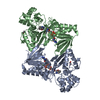
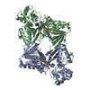
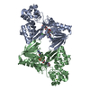
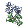
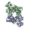
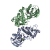
 CHROMOBACTERIUM VIOLACEUM (bacteria)
 X-RAY DIFFRACTION /
  Authors
 Citation
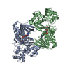
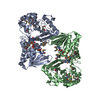
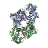
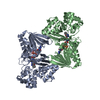
 Structure visualization
 Downloads & links
 Other downloads

 PDBj
PDBj
 Assembly
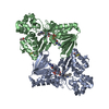

 Mass: 787.566 Da / Num. of mol.: 2 / Source method: obtained synthetically / Formula: C27H35N9O15P2
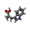
 Mass: 201.221 Da / Num. of mol.: 2 / Source method: obtained synthetically / Formula: C12H11NO2
 Mass: 92.094 Da / Num. of mol.: 1 / Source method: obtained synthetically / Formula: C3H8O3
 Mass: 18.015 Da / Num. of mol.: 297 / Source method: isolated from a natural source / Formula: H2O
 Sample preparation
 / Beamline: 14.1 / Wavelength: 0.918409
  Processing