 Movie
Movie Controller

[English] 日本語
 Yorodumi
- PDB-6fw9: Crystal structure of L-tryptophan oxidase VioA from Chromobacteri... -
+ Open data

- Basic information
| Entry | Database: PDB / ID: 6fw9 | ||||||
|---|---|---|---|---|---|---|---|
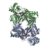
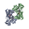
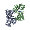
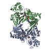
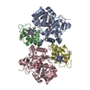
| Title | Crystal structure of L-tryptophan oxidase VioA from Chromobacterium violaceum in complex with 6-Fluoro-L-Tryptophan | ||||||
 Components Components | Flavin-dependent L-tryptophan oxidase VioA | ||||||
 Keywords Keywords | BIOSYNTHETIC PROTEIN / Violacein biosynthesis / L-tryptophan oxidase | ||||||
| Function / homology |  Function and homology information Function and homology information7-chloro-L-tryptophan oxidase / antibiotic biosynthetic process / oxidoreductase activity / metal ion binding Similarity search - Function | ||||||
| Biological species |  Chromobacterium violaceum ATCC 12472 (bacteria) Chromobacterium violaceum ATCC 12472 (bacteria) | ||||||
| Method |  X-RAY DIFFRACTION / SYNCHROTRON / MOLECULAR REPLACEMENT / Resolution: 2.739 Å X-RAY DIFFRACTION / SYNCHROTRON / MOLECULAR REPLACEMENT / Resolution: 2.739 Å | ||||||
 Authors Authors | Lai, H.E. / Morgan, M. / Moore, S. / Freemont, P. | ||||||
| Funding support |  United Kingdom, 1items United Kingdom, 1items
| ||||||
 Citation Citation | Journal: Biorxiv / Year: 2019 Title: A GenoChemetic strategy for derivatization of the violacein natural product scaffold Authors: Lai, H.E. / Obled, A.M.C. / Chee, S.M. / Morgan, R.M. / Sharma, S.V. / Moore, S.J. / Polizzi, K.M. / Goss, R.J.M. / Freemont, P.S. | ||||||
| History |
|
- Structure visualization
| Structure viewer | Molecule: MolmilJmol/JSmol |
|---|
- Downloads & links
-Download
| PDBx/mmCIF format | 6fw9.cif.gz | 182.1 KB | Display | PDBx/mmCIF format |
|---|---|---|---|---|
| PDB format | pdb6fw9.ent.gz | 142 KB | Display | PDB format |
| PDBx/mmJSON format | 6fw9.json.gz | Tree view | PDBx/mmJSON format | |
| Others |  Other downloads Other downloads |
-Validation report
| Arichive directory | https://data.pdbj.org/pub/pdb/validation_reports/fw/6fw9ftp://data.pdbj.org/pub/pdb/validation_reports/fw/6fw9 | HTTPS FTP |
|---|
-Related structure data
| Related structure data |  6fw7C  6fw8C  6fwaC  6ese S: Starting model for refinement C: citing same article ( |
|---|---|
| Similar structure data |
-Links
 PDBj
PDBj
- Assembly
| Deposited unit | 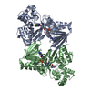
| ||||||||
|---|---|---|---|---|---|---|---|---|---|
| 1 |
| ||||||||
| Unit cell |
|
-Components
| #1: Protein | Mass: 46680.980 Da / Num. of mol.: 2 Source method: isolated from a genetically manipulated source Source: (gene. exp.) Chromobacterium violaceum ATCC 12472 (bacteria)Gene: vioA, CV_3274 / Production host: #2: Chemical |   Mass: 785.550 Da / Num. of mol.: 2 / Source method: obtained synthetically / Formula: C27H33N9O15P2 / Comment: FAD*YM Mass: 785.550 Da / Num. of mol.: 2 / Source method: obtained synthetically / Formula: C27H33N9O15P2 / Comment: FAD*YM#3: Chemical |   Type: L-peptide linking / Mass: 222.216 Da / Num. of mol.: 2 / Source method: obtained synthetically / Formula: C11H11FN2O2 Type: L-peptide linking / Mass: 222.216 Da / Num. of mol.: 2 / Source method: obtained synthetically / Formula: C11H11FN2O2#4: Chemical |   Mass: 24.305 Da / Num. of mol.: 2 / Source method: obtained synthetically / Formula: Mg Mass: 24.305 Da / Num. of mol.: 2 / Source method: obtained synthetically / Formula: Mg#5: Water | ChemComp-HOH / |  Mass: 18.015 Da / Num. of mol.: 118 / Source method: isolated from a natural source / Formula: H2O Mass: 18.015 Da / Num. of mol.: 118 / Source method: isolated from a natural source / Formula: H2OHas protein modification | Y | |
|---|
-Experimental details
-Experiment
| Experiment | Method: X-RAY DIFFRACTION / Number of used crystals: 1 |
|---|
- Sample preparation
| Crystal | Density Matthews: 3.37 Å3/Da / Density % sol: 63.53 % |
|---|---|
| Crystal grow | Temperature: 293 K / Method: vapor diffusion, sitting drop / pH: 7.5 Details: 100 mM HEPES pH 7.5, 9% PEG 8000 and 9% ethylene glycol |
-Data collection
| Diffraction | Mean temperature: 100 K |
|---|---|
| Diffraction source | Source: SYNCHROTRON / Site: Diamond / Beamline: I03 / Wavelength: 0.98 Å |
| Detector | Type: DECTRIS PILATUS3 6M / Detector: PIXEL / Date: Dec 16, 2017 |
| Radiation | Protocol: SINGLE WAVELENGTH / Monochromatic (M) / Laue (L): M / Scattering type: x-ray |
| Radiation wavelength | Wavelength: 0.98 Å / Relative weight: 1 |
| Reflection | Resolution: 2.739→63.86 Å / Num. obs: 33026 / % possible obs: 99 % / Redundancy: 6.4 % / CC1/2: 0.97 / Rmerge(I) obs: 0.1456 / Rrim(I) all: 0.1587 / Net I/σ(I): 8.05 |
| Reflection shell | Resolution: 2.739→2.828 Å / Redundancy: 6.3 % / Num. unique obs: 3237 / % possible all: 100 |
- Processing
| Software |
| ||||||||||||||||||||||||||||||||||||||||||||||||||||||||||||||||||||||||||||||||||||
|---|---|---|---|---|---|---|---|---|---|---|---|---|---|---|---|---|---|---|---|---|---|---|---|---|---|---|---|---|---|---|---|---|---|---|---|---|---|---|---|---|---|---|---|---|---|---|---|---|---|---|---|---|---|---|---|---|---|---|---|---|---|---|---|---|---|---|---|---|---|---|---|---|---|---|---|---|---|---|---|---|---|---|---|---|---|
| Refinement | Method to determine structure: MOLECULAR REPLACEMENT Starting model: 6ESE 6ese Resolution: 2.739→63.857 Å / SU ML: 0.42 / Cross valid method: FREE R-VALUE / σ(F): 1.33 / Phase error: 30.5 / Stereochemistry target values: ML
| ||||||||||||||||||||||||||||||||||||||||||||||||||||||||||||||||||||||||||||||||||||
| Solvent computation | Shrinkage radii: 0.9 Å / VDW probe radii: 1.11 Å / Solvent model: FLAT BULK SOLVENT MODEL | ||||||||||||||||||||||||||||||||||||||||||||||||||||||||||||||||||||||||||||||||||||
| Refinement step | Cycle: LAST / Resolution: 2.739→63.857 Å
| ||||||||||||||||||||||||||||||||||||||||||||||||||||||||||||||||||||||||||||||||||||
| Refine LS restraints |
| ||||||||||||||||||||||||||||||||||||||||||||||||||||||||||||||||||||||||||||||||||||
| LS refinement shell |
|
