 Movie
Movie Controller

[English] 日本語
 Yorodumi
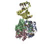
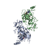
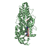
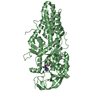
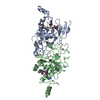
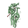
- PDB-5g0j: Crystal structure of Danio rerio HDAC6 CD1 and CD2 (linker intact... -
+ Open data

- Basic information
| Entry | Database: PDB / ID: 5g0j | ||||||
|---|---|---|---|---|---|---|---|
| Title | Crystal structure of Danio rerio HDAC6 CD1 and CD2 (linker intact) in complex with Nexturastat A | ||||||
 Components Components | HDAC6 | ||||||
 Keywords Keywords | CELL CYCLE / HISTONE / HISTONE DEACETYLASE | ||||||
| Function / homology |  Function and homology information Function and homology informationAggrephagy / negative regulation of cellular component organization / positive regulation of cellular component organization / deacetylase activity / tubulin deacetylase activity / mitochondrion localization / definitive hemopoiesis / regulation of microtubule-based process / protein lysine deacetylase activity / potassium ion binding ...Aggrephagy / negative regulation of cellular component organization / positive regulation of cellular component organization / deacetylase activity / tubulin deacetylase activity / mitochondrion localization / definitive hemopoiesis / regulation of microtubule-based process / protein lysine deacetylase activity / potassium ion binding / response to stress / hematopoietic progenitor cell differentiation / swimming behavior / transferase activity / chromatin organization / actin binding / angiogenesis / perikaryon / axon / centrosome / dendrite / zinc ion binding / nucleus / cytosol / cytoplasm Similarity search - Function | ||||||
| Biological species |  | ||||||
| Method |  X-RAY DIFFRACTION / SYNCHROTRON / MOLECULAR REPLACEMENT / Resolution: 2.88 Å X-RAY DIFFRACTION / SYNCHROTRON / MOLECULAR REPLACEMENT / Resolution: 2.88 Å | ||||||
 Authors Authors | Miyake, Y. / Keusch, J.J. / Wang, L. / Saito, M. / Hess, D. / Wang, X. / Melancon, B.J. / Helquist, P. / Gut, H. / Matthias, P. | ||||||
 Citation Citation | Journal: Nat.Chem.Biol. / Year: 2016 Title: Structural Insights Into Hdac6 Tubulin Deacetylation and its Selective Inhibition Authors: Miyake, Y. / Keusch, J.J. / Wang, L. / Saito, M. / Hess, D. / Wang, X. / Melancon, B.J. / Helquist, P. / Gut, H. / Matthias, P. | ||||||
| History |
|
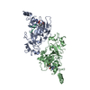
- Structure visualization
| Structure viewer | Molecule: MolmilJmol/JSmol |
|---|
- Downloads & links
-Download
| PDBx/mmCIF format | 5g0j.cif.gz | 167.4 KB | Display | PDBx/mmCIF format |
|---|---|---|---|---|
| PDB format | pdb5g0j.ent.gz | 129.4 KB | Display | PDB format |
| PDBx/mmJSON format | 5g0j.json.gz | Tree view | PDBx/mmJSON format | |
| Others |  Other downloads Other downloads |
-Validation report
| Arichive directory | https://data.pdbj.org/pub/pdb/validation_reports/g0/5g0jftp://data.pdbj.org/pub/pdb/validation_reports/g0/5g0j | HTTPS FTP |
|---|
-Related structure data
| Related structure data |  5g0fC  5g0gSC  5g0hSC  5g0iC C: citing same article ( S: Starting model for refinement |
|---|---|
| Similar structure data |

-Links
 PDBj
PDBj
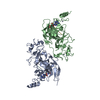
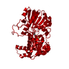
- Assembly
| Deposited unit | 
| ||||||||
|---|---|---|---|---|---|---|---|---|---|
| 1 |
| ||||||||
| Unit cell |
|
-Components
-Protein , 1 types, 1 molecules A
| #1: Protein | Mass: 87514.406 Da / Num. of mol.: 1 / Fragment: CATALYTIC DOMAIN 1 AND 2, UNP RESIDUES 40-831 Source method: isolated from a genetically manipulated source Source: (gene. exp.)   SPODOPTERA FRUGIPERDA (fall armyworm) / References: UniProt: F8W4B7 SPODOPTERA FRUGIPERDA (fall armyworm) / References: UniProt: F8W4B7 |
|---|
-Non-polymers , 5 types, 171 molecules 


| #2: Chemical |  Mass: 35.453 Da / Num. of mol.: 2 / Source method: obtained synthetically / Formula: Cl Mass: 35.453 Da / Num. of mol.: 2 / Source method: obtained synthetically / Formula: Cl#3: Chemical | ChemComp-ZN / |  Mass: 65.409 Da / Num. of mol.: 1 / Source method: obtained synthetically / Formula: Zn Mass: 65.409 Da / Num. of mol.: 1 / Source method: obtained synthetically / Formula: Zn#4: Chemical | ChemComp-K /  Mass: 39.098 Da / Num. of mol.: 5 / Source method: obtained synthetically / Formula: K Mass: 39.098 Da / Num. of mol.: 5 / Source method: obtained synthetically / Formula: K#5: Chemical | ChemComp-N4R / |  Mass: 341.404 Da / Num. of mol.: 1 / Source method: obtained synthetically / Formula: C19H23N3O3 Mass: 341.404 Da / Num. of mol.: 1 / Source method: obtained synthetically / Formula: C19H23N3O3#6: Water | ChemComp-HOH / | Mass: 18.015 Da / Num. of mol.: 162 / Source method: isolated from a natural source / Formula: H2O |
|---|
-Experimental details
-Experiment
| Experiment | Method: X-RAY DIFFRACTION / Number of used crystals: 1 |
|---|
- Sample preparation
| Crystal | Density Matthews: 5.6 Å3/Da / Density % sol: 78 % / Description: NONE |
|---|---|
| Crystal grow | Details: 3.3M SODIUM FORMATE, 17% GLYCEROL, 0.1M TRIS PH 7.5 |
-Data collection
| Diffraction | Mean temperature: 100 K |
|---|---|
| Diffraction source | Source: SYNCHROTRON / Site: SLS  / Beamline: X10SA / Wavelength: 1 / Beamline: X10SA / Wavelength: 1 |
| Detector | Type: DECTRIS PILATUS 6M / Detector: PIXEL / Details: MIRRORS |
| Radiation | Protocol: SINGLE WAVELENGTH / Monochromatic (M) / Laue (L): M / Scattering type: x-ray |
| Radiation wavelength | Wavelength: 1 Å / Relative weight: 1 |
| Reflection | Resolution: 2.88→162.14 Å / Num. obs: 47289 / % possible obs: 100 % / Observed criterion σ(I): -3 / Redundancy: 11.2 % / Biso Wilson estimate: 88.96 Å2 / Rmerge(I) obs: 0.08 / Net I/σ(I): 20.4 |
| Reflection shell | Resolution: 2.88→2.89 Å / Redundancy: 11.3 % / Rmerge(I) obs: 0.79 / Mean I/σ(I) obs: 2.4 / % possible all: 100 |
- Processing
| Software |
| ||||||||||||||||||||||||||||||||||||||||||||||||||||||||||||||||||||||||||||||||||||||||||||||||||||||||||||||||||
|---|---|---|---|---|---|---|---|---|---|---|---|---|---|---|---|---|---|---|---|---|---|---|---|---|---|---|---|---|---|---|---|---|---|---|---|---|---|---|---|---|---|---|---|---|---|---|---|---|---|---|---|---|---|---|---|---|---|---|---|---|---|---|---|---|---|---|---|---|---|---|---|---|---|---|---|---|---|---|---|---|---|---|---|---|---|---|---|---|---|---|---|---|---|---|---|---|---|---|---|---|---|---|---|---|---|---|---|---|---|---|---|---|---|---|---|
| Refinement | Method to determine structure: MOLECULAR REPLACEMENT Starting model: PDB ENTRIES 5G0G AND 5G0H Resolution: 2.88→50 Å / Cor.coef. Fo:Fc: 0.898 / Cor.coef. Fo:Fc free: 0.8705 / SU R Cruickshank DPI: 0.259 / Cross valid method: THROUGHOUT / σ(F): 0 / SU R Blow DPI: 0.254 / SU Rfree Blow DPI: 0.216 / SU Rfree Cruickshank DPI: 0.22 Details: DIFFRACTION DATA ARE ANISOTROPIC. STRUCTURAL MODELING WAS CARRIED OUT USING MAP SHARPENING
| ||||||||||||||||||||||||||||||||||||||||||||||||||||||||||||||||||||||||||||||||||||||||||||||||||||||||||||||||||
| Displacement parameters | Biso mean: 90.06 Å2
| ||||||||||||||||||||||||||||||||||||||||||||||||||||||||||||||||||||||||||||||||||||||||||||||||||||||||||||||||||
| Refine analyze | Luzzati coordinate error obs: 0.383 Å | ||||||||||||||||||||||||||||||||||||||||||||||||||||||||||||||||||||||||||||||||||||||||||||||||||||||||||||||||||
| Refinement step | Cycle: LAST / Resolution: 2.88→50 Å
| ||||||||||||||||||||||||||||||||||||||||||||||||||||||||||||||||||||||||||||||||||||||||||||||||||||||||||||||||||
| Refine LS restraints |
| ||||||||||||||||||||||||||||||||||||||||||||||||||||||||||||||||||||||||||||||||||||||||||||||||||||||||||||||||||
| LS refinement shell | Resolution: 2.88→2.96 Å / Total num. of bins used: 20
|
