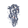
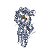
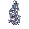
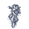
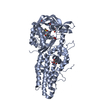
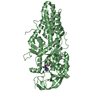
Entry Database : PDB / ID : 5j5aTitle Trypanosoma brucei methionyl-tRNA synthetase in complex with inhibitor (Chem 70786556) Methionyl-tRNA synthetase, putative Keywords / / / / / Function / homology Function Domain/homology Component
/ / / / / / / / / / / / / / / / / / / / / / / / / / / / / / / / / / / Biological species Trypanosoma brucei brucei (eukaryote)Method / / Resolution : 2.7 Å Authors Barros-Alvarez, X. / Koh, C.Y. / Hol, W.G.J. Journal : Eur J Med Chem / Year : 2016Title : Structure-guided design of novel Trypanosoma brucei Methionyl-tRNA synthetase inhibitors.Authors : Huang, W. / Zhang, Z. / Barros-Alvarez, X. / Koh, C.Y. / Ranade, R.M. / Gillespie, J.R. / Creason, S.A. / Shibata, S. / Verlinde, C.L. / Hol, W.G. / Buckner, F.S. / Fan, E. History Deposition Apr 1, 2016 Deposition site / Processing site Revision 1.0 Jan 25, 2017 Provider / Type Revision 1.1 Mar 6, 2024 Group / Database references / Refinement descriptionCategory chem_comp_atom / chem_comp_bond ... chem_comp_atom / chem_comp_bond / database_2 / struct_ncs_dom_lim Item _database_2.pdbx_DOI / _database_2.pdbx_database_accession ... _database_2.pdbx_DOI / _database_2.pdbx_database_accession / _struct_ncs_dom_lim.beg_auth_comp_id / _struct_ncs_dom_lim.beg_label_asym_id / _struct_ncs_dom_lim.beg_label_comp_id / _struct_ncs_dom_lim.beg_label_seq_id / _struct_ncs_dom_lim.end_auth_comp_id / _struct_ncs_dom_lim.end_label_asym_id / _struct_ncs_dom_lim.end_label_comp_id / _struct_ncs_dom_lim.end_label_seq_id
Show all Show less
 Movie
Movie Controller

 Yorodumi
 Open data
 Basic information
 Components
 Keywords
 Function and homology information
 X-RAY DIFFRACTION /
  Authors
 Citation
 Structure visualization
 Downloads & links
 Other downloads


 PDBj
PDBj
 Assembly
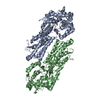


 Type: L-peptide linking / Mass: 149.211 Da / Num. of mol.: 1 / Source method: obtained synthetically / Formula: C5H11NO2S
 Mass: 359.421 Da / Num. of mol.: 1 / Source method: obtained synthetically / Formula: C22H21N3O2
 Mass: 18.015 Da / Num. of mol.: 249 / Source method: isolated from a natural source / Formula: H2O
 Sample preparation
 Processing