- PDB-2qs7: CRYSTAL STRUCTURE OF a putative oxidoreductase of the DsrE/DsrF-l... -
+
Open data
ID or keywords:
Loading...
-
Basic information
Entry
Database: PDB / ID: 2qs7
Title
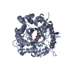
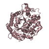
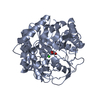
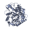
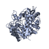
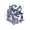
CRYSTAL STRUCTURE OF a putative oxidoreductase of the DsrE/DsrF-like family (SSO1126) FROM SULFOLOBUS SOLFATARICUS P2 AT 2.09 A RESOLUTION
Components
Uncharacterized protein
Keywords
OXIDOREDUCTASE / PUTATIVE OXIDOREDUCTASE OF THE DSRE/DSRF-LIKE FAMILY / STRUCTURAL GENOMICS / JOINT CENTER FOR STRUCTURAL GENOMICS / JCSG / PROTEIN STRUCTURE INITIATIVE / PSI-2
Function / homology
DsrE2-like family / DsrE/DsrF/DrsH-like family / DsrEFH-like / DsrEFH-like / Hypothetical Protein Ychn; Chain: A, / 3-Layer(aba) Sandwich / Alpha Beta / Peroxiredoxin
Function and homology information
Biological species
Sulfolobus solfataricus P2 (archaea)
Method
X-RAY DIFFRACTION / SYNCHROTRON / MAD / Resolution: 2.09 Å
BIOMOLECULE: 1, 2 SEE REMARK 350 FOR THE AUTHOR PROVIDED AND PROGRAM GENERATED ASSEMBLY ... BIOMOLECULE: 1, 2 SEE REMARK 350 FOR THE AUTHOR PROVIDED AND PROGRAM GENERATED ASSEMBLY INFORMATION FOR THE STRUCTURE IN THIS ENTRY. THE REMARK MAY ALSO PROVIDE INFORMATION ON BURIED SURFACE AREA. SIZE EXCLUSION CHROMATOGRAPHY WITH STATIC LIGHT SCATTERING SUPPORTS THE ASSIGNMENT OF A TRIMER AS A SIGNIFICANT OLIGOMERIZATION STATE.
Remark 999
SEQUENCE THE CONSTRUCT WAS EXPRESSED WITH A PURIFICATION TAG MGSDKIHHHHHHENLYFQG. THE TAG WAS ... SEQUENCE THE CONSTRUCT WAS EXPRESSED WITH A PURIFICATION TAG MGSDKIHHHHHHENLYFQG. THE TAG WAS REMOVED WITH TEV PROTEASE LEAVING ONLY A GLYCINE (0) FOLLOWED BY THE TARGET SEQUENCE.
Resolution: 2.09→48.737 Å / Num. obs: 42142 / % possible obs: 98.9 % / Observed criterion σ(I): -3 / Biso Wilson estimate: 48.55 Å2 / Rmerge(I) obs: 0.066 / Net I/σ(I): 21.12
Reflection shell
Resolution (Å)
Rmerge(I) obs
Mean I/σ(I) obs
Num. measured obs
Num. unique obs
Diffraction-ID
% possible all
2.09-2.16
0.801
2.24
19998
3267
1,2
88.8
2.16-2.25
0.617
3.3
25974
4171
1,2
95
2.25-2.35
0.442
4.6
24847
3996
1,2
96.3
2.35-2.48
0.335
6.2
26770
4294
1,2
97.3
2.48-2.63
0.358
9.6
36054
4029
1,2
98.3
2.63-2.83
0.256
15.4
46326
4143
1,2
99
2.83-3.12
0.149
24.6
51040
4333
1,2
99.5
3.12-3.57
0.085
38
49524
4247
1,2
99.6
3.57-48.737
0.056
50.6
49239
4299
1,2
99.9
-
Phasing
Phasing
Method: MAD
-
Processing
Software
Name
Version
Classification
NB
REFMAC
5.2.0019
refinement
PHENIX
refinement
SHELX
phasing
MolProbity
3beta29
modelbuilding
XSCALE
datascaling
PDB_EXTRACT
3
dataextraction
MAR345
CCD
datacollection
XDS
datareduction
SHELXD
phasing
autoSHARP
phasing
Refinement
Method to determine structure: MAD / Resolution: 2.09→48.737 Å / Cor.coef. Fo:Fc: 0.97 / Cor.coef. Fo:Fc free: 0.96 / SU B: 9.09 / SU ML: 0.117 / TLS residual ADP flag: LIKELY RESIDUAL / Cross valid method: THROUGHOUT / σ(F): 0 / ESU R: 0.167 / ESU R Free: 0.144 Stereochemistry target values: MAXIMUM LIKELIHOOD WITH PHASES Details: 1. HYDROGENS HAVE BEEN ADDED IN THE RIDING POSITIONS. 2. ATOM RECORDS CONTAIN RESIDUAL B FACTORS ONLY. 3. A MET-INHIBITION PROTOCOL WAS USED FOR SELENOMETHIONINE INCORPORATION DURING PROTEIN ...Details: 1. HYDROGENS HAVE BEEN ADDED IN THE RIDING POSITIONS. 2. ATOM RECORDS CONTAIN RESIDUAL B FACTORS ONLY. 3. A MET-INHIBITION PROTOCOL WAS USED FOR SELENOMETHIONINE INCORPORATION DURING PROTEIN EXPRESSION. THE OCCUPANCY OF THE SE ATOMS IN THE MSE RESIDUES WAS REDUCED TO 0.75 FOR THE REDUCED SCATTERING POWER DUE TO PARTIAL S-MET INCORPORATION. 4. CL IONS FROM THE CRYSTALLIZATION SOLUTION ARE MODELED. HEPES (EPE) MOLECULES WERE MODELED BASED ON DENSITY. 5. WATERS 6,7,8,10 COULD BE LIGHT METAL IONS SUCH AS MAGNESIUM. 6. DENSITIES FOR RESIDUES 59-61 ARE POOR.
Rfactor
Num. reflection
% reflection
Selection details
Rfree
0.203
2098
5 %
RANDOM
Rwork
0.176
-
-
-
all
0.177
-
-
-
obs
0.177
42142
99.62 %
-
Solvent computation
Ion probe radii: 0.8 Å / Shrinkage radii: 0.8 Å / VDW probe radii: 1.2 Å / Solvent model: BABINET MODEL WITH MASK
Displacement parameters
Biso mean: 49.899 Å2
Baniso -1
Baniso -2
Baniso -3
1-
0.62 Å2
0.31 Å2
0 Å2
2-
-
0.62 Å2
0 Å2
3-
-
-
-0.94 Å2
Refinement step
Cycle: LAST / Resolution: 2.09→48.737 Å
Protein
Nucleic acid
Ligand
Solvent
Total
Num. atoms
4222
0
33
85
4340
Refine LS restraints
Refine-ID
Type
Dev ideal
Dev ideal target
Number
X-RAY DIFFRACTION
r_bond_refined_d
0.017
0.022
4451
X-RAY DIFFRACTION
r_bond_other_d
0.003
0.02
3014
X-RAY DIFFRACTION
r_angle_refined_deg
1.562
1.965
6028
X-RAY DIFFRACTION
r_angle_other_deg
1.048
3
7401
X-RAY DIFFRACTION
r_dihedral_angle_1_deg
3.122
5
574
X-RAY DIFFRACTION
r_dihedral_angle_2_deg
31.706
24.471
170
X-RAY DIFFRACTION
r_dihedral_angle_3_deg
13.377
15
778
X-RAY DIFFRACTION
r_dihedral_angle_4_deg
10.252
15
12
X-RAY DIFFRACTION
r_chiral_restr
0.101
0.2
677
X-RAY DIFFRACTION
r_gen_planes_refined
0.006
0.02
4897
X-RAY DIFFRACTION
r_gen_planes_other
0.002
0.02
915
X-RAY DIFFRACTION
r_nbd_refined
0.231
0.2
902
X-RAY DIFFRACTION
r_nbd_other
0.187
0.2
2898
X-RAY DIFFRACTION
r_nbtor_refined
0.191
0.2
2214
X-RAY DIFFRACTION
r_nbtor_other
0.091
0.2
2248
X-RAY DIFFRACTION
r_xyhbond_nbd_refined
0.202
0.2
103
X-RAY DIFFRACTION
r_symmetry_vdw_refined
0.13
0.2
21
X-RAY DIFFRACTION
r_symmetry_vdw_other
0.297
0.2
44
X-RAY DIFFRACTION
r_symmetry_hbond_refined
0.333
0.2
11
X-RAY DIFFRACTION
r_mcbond_it
1.977
3
3012
X-RAY DIFFRACTION
r_mcbond_other
0.323
3
1137
X-RAY DIFFRACTION
r_mcangle_it
2.695
5
4474
X-RAY DIFFRACTION
r_scbond_it
5.237
8
1872
X-RAY DIFFRACTION
r_scangle_it
6.971
11
1541
Refine LS restraints NCS
Ens-ID: 1 / Refine-ID: X-RAY DIFFRACTION
Dom-ID
Auth asym-ID
Number
Type
Rms dev position (Å)
Weight position
1
A
802
TIGHTPOSITIONAL
0.06
0.05
2
B
802
TIGHTPOSITIONAL
0.04
0.05
3
C
802
TIGHTPOSITIONAL
0.06
0.05
4
D
802
TIGHTPOSITIONAL
0.05
0.05
1
A
847
MEDIUMPOSITIONAL
0.34
0.5
2
B
847
MEDIUMPOSITIONAL
0.42
0.5
3
C
847
MEDIUMPOSITIONAL
0.4
0.5
4
D
847
MEDIUMPOSITIONAL
0.35
0.5
1
A
802
TIGHTTHERMAL
0.17
0.5
2
B
802
TIGHTTHERMAL
0.15
0.5
3
C
802
TIGHTTHERMAL
0.16
0.5
4
D
802
TIGHTTHERMAL
0.19
0.5
1
A
847
MEDIUMTHERMAL
1.17
2
2
B
847
MEDIUMTHERMAL
0.96
2
3
C
847
MEDIUMTHERMAL
1.17
2
4
D
847
MEDIUMTHERMAL
1.18
2
LS refinement shell
Resolution: 2.09→2.149 Å / Total num. of bins used: 20
Rfactor
Num. reflection
% reflection
Rfree
0.333
148
-
Rwork
0.269
2813
-
obs
-
2961
95.64 %
Refinement TLS params.
Method: refined / Refine-ID: X-RAY DIFFRACTION
ID
L11 (°2)
L12 (°2)
L13 (°2)
L22 (°2)
L23 (°2)
L33 (°2)
S11 (Å °)
S12 (Å °)
S13 (Å °)
S21 (Å °)
S22 (Å °)
S23 (Å °)
S31 (Å °)
S32 (Å °)
S33 (Å °)
T11 (Å2)
T12 (Å2)
T13 (Å2)
T22 (Å2)
T23 (Å2)
T33 (Å2)
Origin x (Å)
Origin y (Å)
Origin z (Å)
1
2.817
0.747
0.2113
6.203
0.4786
4.1435
-0.1011
0.1313
0.0472
-0.5629
-0.1517
1.0288
-0.0619
-0.8241
0.2528
-0.2306
-0.0567
-0.1558
0.097
-0.1055
-0.024
-36.6354
-6.1981
-24.2314
2
4.8558
-0.6234
0.5608
4.9436
-0.0251
5.14
-0.0014
0.1655
-0.9176
-0.2155
-0.2134
0.0438
1.0647
0.0048
0.2147
0.0002
-0.0958
0.0529
-0.1535
-0.0303
0.0407
-23.8253
-25.8199
-20.8535
3
2.9349
-1.1249
0.0916
4.8627
1.2464
4.4857
-0.1707
-0.5571
-0.7552
0.7864
-0.2022
0.795
0.9065
-0.9393
0.3729
0.0254
-0.2782
0.186
0.2306
-0.0114
0.0437
-37.4425
-17.2914
-3.4358
4
4.6544
0.1437
-0.1172
3.9115
-0.3533
2.5236
-0.1001
0.6061
-0.7183
-0.6441
-0.004
0.0695
0.4258
0.0034
0.1041
-0.0855
-0.0102
0.0343
-0.1586
-0.1333
-0.2064
-0.6803
-13.5808
-41.6236
Refinement TLS group
ID
Refine-ID
Refine TLS-ID
Auth asym-ID
Label asym-ID
Auth seq-ID
Label seq-ID
1
X-RAY DIFFRACTION
1
A
A
6 - 143
7 - 144
2
X-RAY DIFFRACTION
2
B
B
6 - 143
7 - 144
3
X-RAY DIFFRACTION
3
C
C
6 - 143
7 - 144
4
X-RAY DIFFRACTION
4
D
D
6 - 143
7 - 144
+
About Yorodumi
-
News
-
Feb 9, 2022. New format data for meta-information of EMDB entries
New format data for meta-information of EMDB entries
Version 3 of the EMDB header file is now the official format.
The previous official version 1.9 will be removed from the archive.
In the structure databanks used in Yorodumi, some data are registered as the other names, "COVID-19 virus" and "2019-nCoV". Here are the details of the virus and the list of structure data.
Jan 31, 2019. EMDB accession codes are about to change! (news from PDBe EMDB page)
EMDB accession codes are about to change! (news from PDBe EMDB page)
The allocation of 4 digits for EMDB accession codes will soon come to an end. Whilst these codes will remain in use, new EMDB accession codes will include an additional digit and will expand incrementally as the available range of codes is exhausted. The current 4-digit format prefixed with “EMD-” (i.e. EMD-XXXX) will advance to a 5-digit format (i.e. EMD-XXXXX), and so on. It is currently estimated that the 4-digit codes will be depleted around Spring 2019, at which point the 5-digit format will come into force.
The EM Navigator/Yorodumi systems omit the EMD- prefix.
Related info.:Q: What is EMD? / ID/Accession-code notation in Yorodumi/EM Navigator
Yorodumi is a browser for structure data from EMDB, PDB, SASBDB, etc.
This page is also the successor to EM Navigator detail page, and also detail information page/front-end page for Omokage search.
The word "yorodu" (or yorozu) is an old Japanese word meaning "ten thousand". "mi" (miru) is to see.
Related info.:EMDB / PDB / SASBDB / Comparison of 3 databanks / Yorodumi Search / Aug 31, 2016. New EM Navigator & Yorodumi / Yorodumi Papers / Jmol/JSmol / Function and homology information / Changes in new EM Navigator and Yorodumi
 Movie
Movie Controller

 Yorodumi
 Open data
 Basic information
 Components
 Keywords
 Function and homology information
 Sulfolobus solfataricus P2 (archaea)
 X-RAY DIFFRACTION /
  Authors
 Citation
 Structure visualization
 Downloads & links
 Other downloads
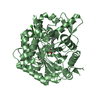


 PDBj
PDBj Assembly
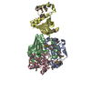


 Mass: 35.453 Da / Num. of mol.: 3 / Source method: obtained synthetically / Formula: Cl
 Mass: 238.305 Da / Num. of mol.: 2 / Source method: obtained synthetically / Formula: C8H18N2O4S / Comment: pH buffer*YM
 Mass: 18.015 Da / Num. of mol.: 85 / Source method: isolated from a natural source / Formula: H2O
 Sample preparation
 Processing